


-
Retinitis pigmentosa
 41yo
41yo 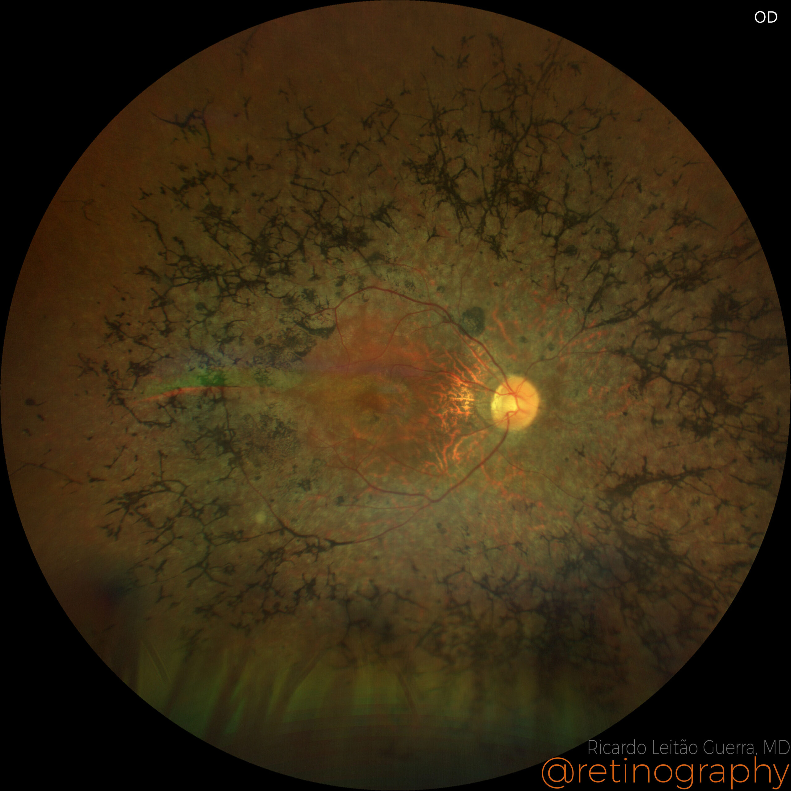 Retinitis pigmentosa
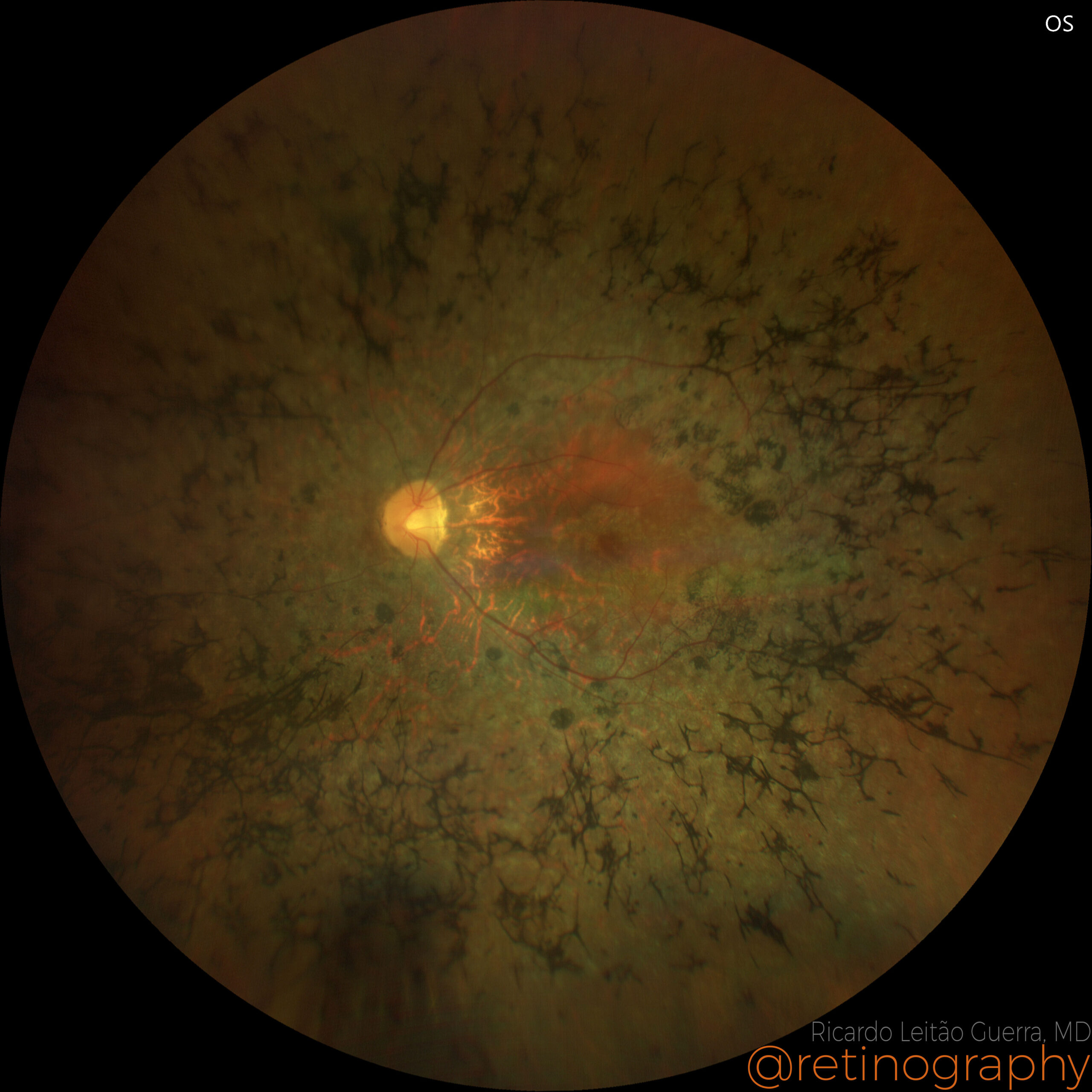 True color
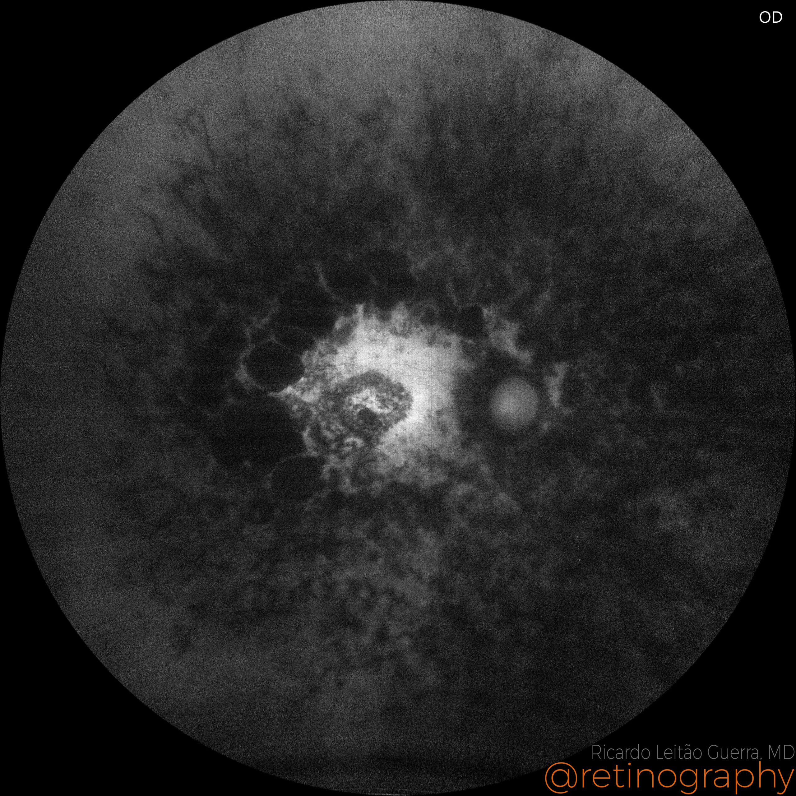 FAF - Green
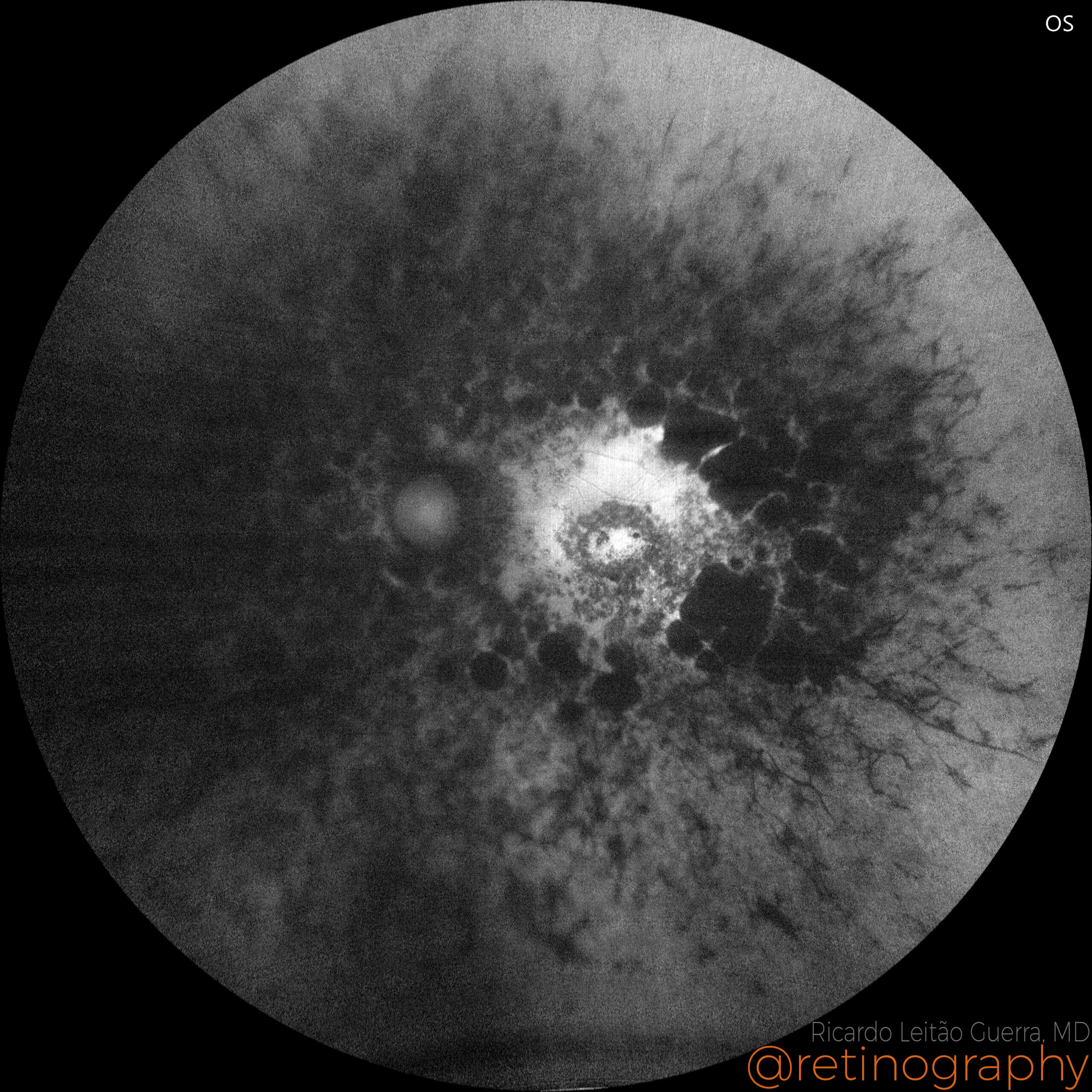 FAF - Green
Retinitis pigmentosa (RP) is a progressive genetic disorder characterized by the degeneration of photoreceptor cells, primarily affecting night and peripheral vision. Fundus autofluorescence (FAF) is a key diagnostic tool in monitoring RP, utilizing the natural fluorescence of lipofuscin in the retinal pigment epithelium (RPE) to detect areas of activity and atrophy. FAF imaging helps identify patterns of progression and areas of retinal degeneration, providing crucial insights for assessing disease severity and potential therapeutic responses.
#retina #oftalmo #ophthalmology #oftalmologia #oftalmología #ophtalmologie #офтальмологія #офтальмология #οφθαλμολογία #retinography2024 #CIRRUS6000 #CLARUS700 #ZEISSRETINAWORKFLOW
Other Cases
-
Proliferative diabetic retinopathy
38yo 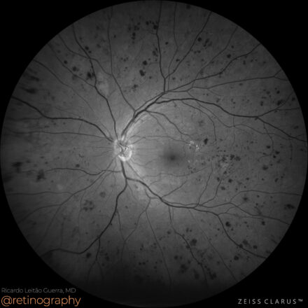
In proliferative diabetic retinopathy (PDR), neovascularization elsewhere (NVE) refers to abnormal blood vessel growth in the midperiphery or periphery of the retina. The green channel enhances contrast, making NVE more visible against the retinal background. Identifying NVE early is crucial for guiding treatment, such as panretinal photocoagulation (PRP) or anti-VEGF […]
-
Proliferative Diabetic Retinopathy
38yo 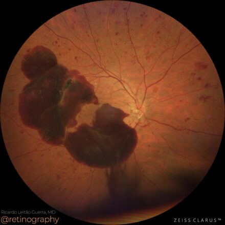
In proliferative diabetic retinopathy (PDR), subhyaloid hemorrhage results from fragile neovascularization bleeding into the preretinal space. The green channel enhances contrast, improving hemorrhage visualization on fundus imaging. Optical Coherence Tomography (OCT) shows a well-demarcated, hyperreflective hemorrhagic pocket between the retina and the posterior hyaloid face, which can affect visual acuity […]
-
AMD: Soft Drusen
 80yo
80yo 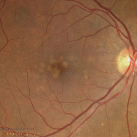
In age-related macular degeneration (AMD), soft confluent drusen appear as yellowish deposits on true color imaging, often coalescing into larger lesions. Optical Coherence Tomography (OCT) reveals these drusen as elevations of the retinal pigment epithelium (RPE) with underlying hyporeflective spaces. The presence of large, confluent drusen increases the risk of […]
-
AMD: Outer retinal tubulations
77yo 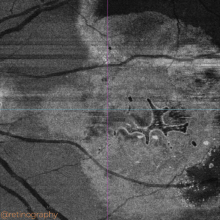
In age-related macular degeneration (AMD), outer retinal tubulations (ORT) can present unique patterns on en-face OCT, often resembling intraretinal fluid (IRF), leading to potential misdiagnosis. In the presented case, en-face imaging shows ORT as hyporreflective interconnected branching networks extensions surrounded by a hyperreflective borders, while dark areas without hyperreflective borders […]
-
Central Serous Chorioretinopathy
40yo 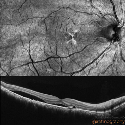
In Central Serous Chorioretinopathy (CSC), serous retinal detachment and serous pigment epithelial detachment (PED) are hallmark findings. Optical Coherence Tomography (OCT) reveals a hyporeflective space beneath the neurosensory retina and PEDs, often associated with focal RPE abnormalities. The leakage site at the RPE can be identified on OCT as a […]
-
Situs inversus: Degenerative myopia
40yo 
In degenerative myopia, situs inversus refers to the tilted insertion of the optic disc, commonly associated with retinal and choroidal changes. Optical Coherence Tomography (OCT) B-scan often shows tilted scans due to the oblique orientation of posterior pole structures. This tilting can distort retinal layer visualization, requiring careful interpretation to […]
-
Polypoidal Choroidal Vasculopathy
79yo 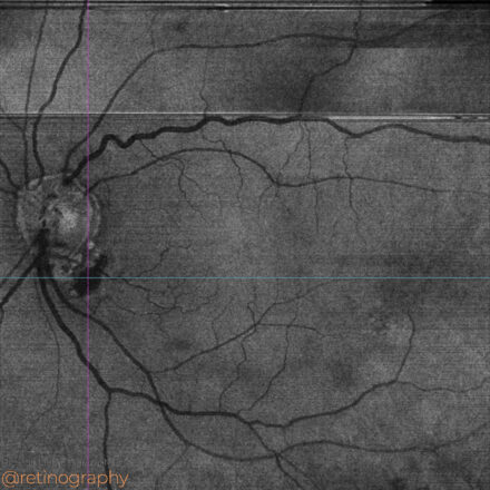
Polypoidal Choroidal Vasculopathy (PCV) should be investigated when subretinal fluid (SRF) is observed adjacent to the optic disc. En-face OCT imaging at the level of the ellipsoid zone (EZ) is a valuable tool for detecting SRF in these cases, providing a detailed view of fluid distribution and its impact on […]
-
AMD: Disciform scar
77yo 
In advanced age-related macular degeneration (AMD), a disciform scar represents the end stage of neovascular AMD. Optical Coherence Tomography (OCT) shows hyperreflective fibrotic tissue replacing normal retinal layers, with possible subretinal fluid or retinal thinning. Fundus autofluorescence (FAF) reveals hypoautofluorescence due to retinal pigment epithelium (RPE) atrophy, with surrounding hyperautofluorescence […]
-
Central Serous Chorioretinopathy
48yo 
In acute Central Serous Chorioretinopathy (CSC), Optical Coherence Tomography (OCT) reveals subretinal fluid as a hyporeflective space between the neurosensory retina and the retinal pigment epithelium (RPE). The absence of significant intraretinal fluid differentiates CSC from other causes of macular edema. OCT is essential for diagnosing, monitoring resolution, and assessing […]
-
Multifocal Best’s vitelliform dystrophy
33yo 
Multifocal Best vitelliform dystrophy is a rare retinal condition characterized by multiple yellowish vitelliform lesions scattered across the posterior pole. Fundus autofluorescence (FAF) shows hyperautofluorescent lesions due to lipofuscin accumulation in the retinal pigment epithelium (RPE). Over time, areas of hypoautofluorescence may appear, indicating RPE atrophy. FAF is crucial for […]
-
Drusen
37yo 
The presence of drusen in a young woman may suggest inherited or systemic conditions rather than typical age-related macular degeneration (AMD). Conditions like familial drusen, basal laminar drusen, or early-onset drusen should be considered. Optical Coherence Tomography (OCT) can reveal their size and location, and fundus autofluorescence (FAF) may show […]
-
Polypoidal choroidal vasculopathy
81yo 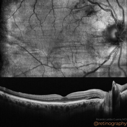
Polypoidal Choroidal Vasculopathy (PCV) should be investigated when subretinal fluid is observed adjacent to the optic disc. Fundus autofluorescence (FAF) is useful in these cases, as it highlights retinal pigment epithelium (RPE) changes. Areas of hyperautofluorescence indicate RPE stress, while hypoautofluorescence suggests atrophy, aiding in the identification of PCV-related abnormalities. […]