


Tag:
Acquired vitelliform lesion
-
Acquired Vitelliform Lesion
Ricardo Leitão Guerra, MD 74yo
74yo  Blue channel
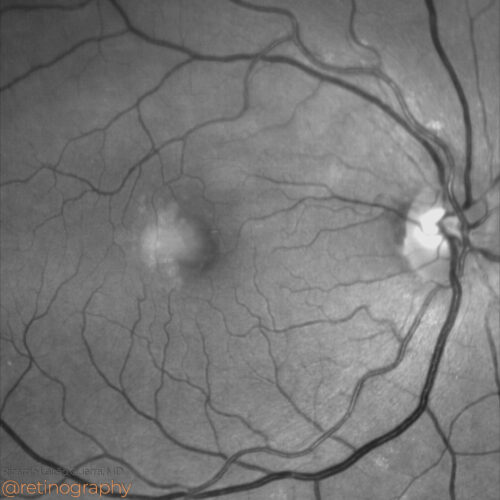 Green Channel
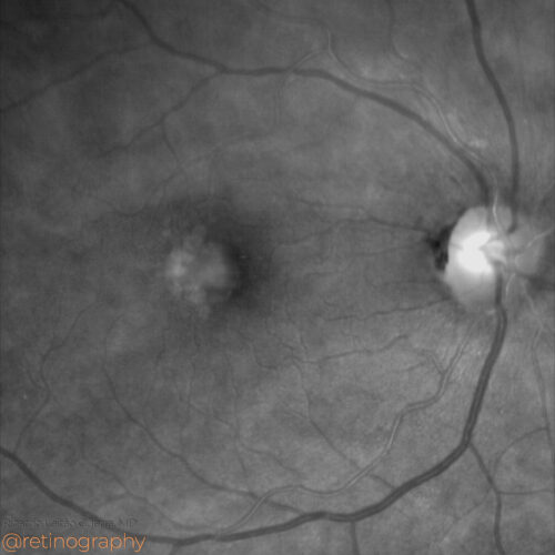 Red channel
 FAF-Green
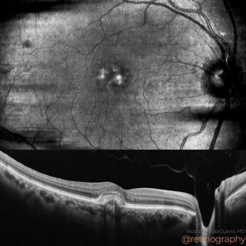 NIR & SD-OCT
Acquired Vitelliform Lesions (AVL) are characterized by subretinal yellowish deposits and can be assessed using fundus autofluorescence (FAF). FAF imaging shows hyperautofluorescence in the ...
Acquired Vitelliform Lesions (AVL) are characterized by subretinal yellowish deposits and can be assessed using fundus autofluorescence (FAF). FAF imaging shows hyperautofluorescence in the ...
Acquired Vitelliform Lesions (AVL) are characterized by subretinal yellowish deposits and can be assessed using fundus autofluorescence (FAF). FAF imaging shows hyperautofluorescence in the lesion area due to lipofuscin accumulation. Over time, hypoautofluorescence may appear as the retinal pigment epithelium (RPE) degenerates, making FAF essential for monitoring disease progression.
#AVL #FAF #Hyperautofluorescence #Lipofuscin #RPE #RetinaImaging #retina #oftalmo #ophthalmology #oftalmologia #oftalmología #ophtalmologie #офтальмологія #офтальмология #οφθαλμολογία #retinography2024 #CIRRUS6000 #CLARUS700 #ZEISSRETINAWORKFLOW
BackRead More -
Acquired Vitelliform Lesion
Ricardo Leitão Guerra, MD 80yo 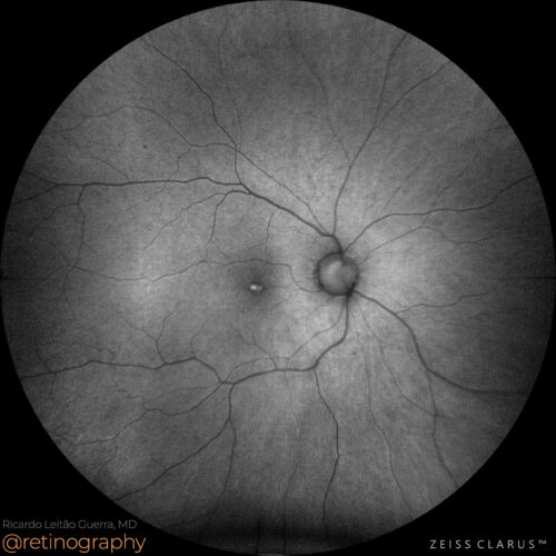 FAF-Green
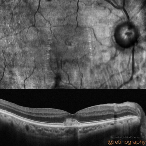 NIR & SD-OCT
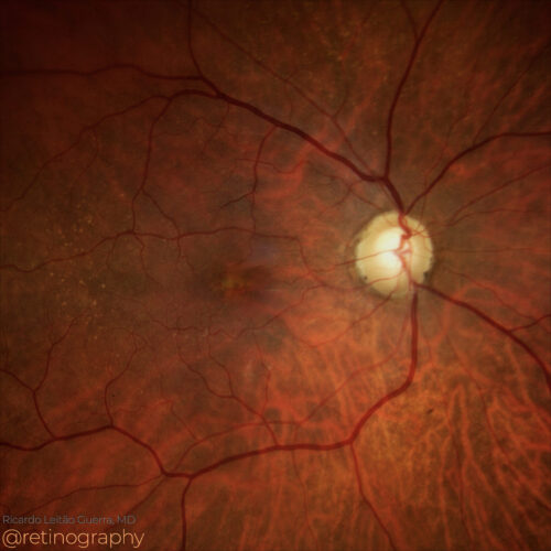 True color
 FAF-Green
Acquired Vitelliform Lesions (AVL) are characterized by subretinal yellowish deposits that can be assessed using multimodal imaging. Fundus autofluorescence (FAF) shows hyperautofluorescence...
Acquired Vitelliform Lesions (AVL) are characterized by subretinal yellowish deposits that can be assessed using multimodal imaging. Fundus autofluorescence (FAF) shows hyperautofluorescence...
Acquired Vitelliform Lesions (AVL) are characterized by subretinal yellowish deposits that can be assessed using multimodal imaging. Fundus autofluorescence (FAF) shows hyperautofluorescence in the area of the lesion due to lipofuscin accumulation. Optical Coherence Tomography (OCT) reveals these deposits as hyperreflective material between the retinal pigment epithelium (RPE) and photoreceptors, helping to monitor progression and assess the impact on retinal structure.
#AVL #VitelliformLesions #FAF #OCT #RetinaImaging #RPE #retina #oftalmo #ophthalmology #oftalmologia #oftalmología #ophtalmologie #офтальмологія #офтальмология #οφθαλμολογία #retinography2024 #CIRRUS6000 #CLARUS700 #ZEISSRETINAWORKFLOW
BackRead More -
Acquired vitelliforme lesion
Ricardo Leitão Guerra, MD 64yo 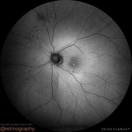 FAF-Green
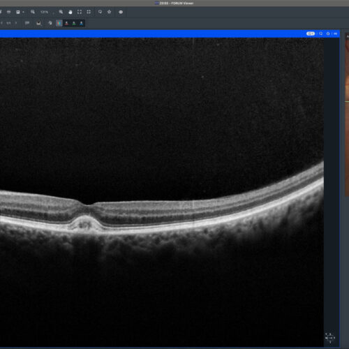 SD-OCT
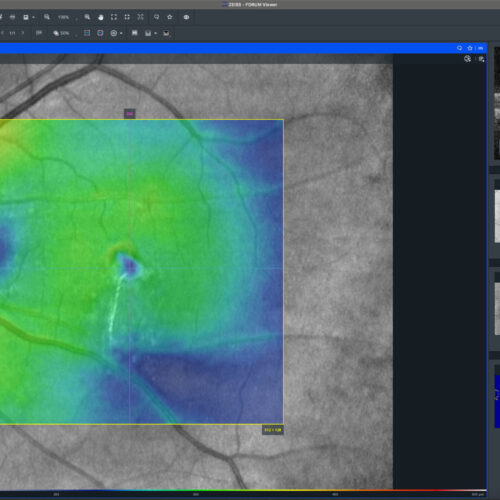 Thickness map
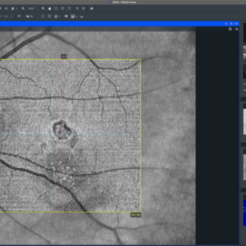 En-face: EZ
Acquired Vitelliform Lesions (AVL) present clinically as yellowish subretinal deposits often seen in conditions like age-related macular degeneration (AMD) and pattern dystrophies. Fundus au...
Acquired Vitelliform Lesions (AVL) present clinically as yellowish subretinal deposits often seen in conditions like age-related macular degeneration (AMD) and pattern dystrophies. Fundus au...
BackRead More -
Acquired vitelliform lesion
Ricardo Leitão Guerra, MD 78yo 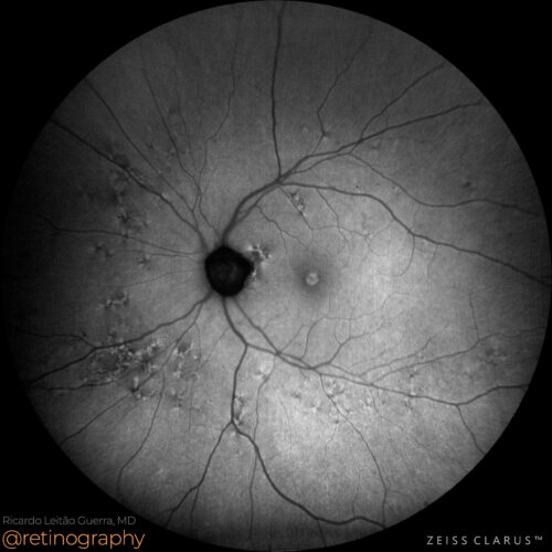 FAF-Green
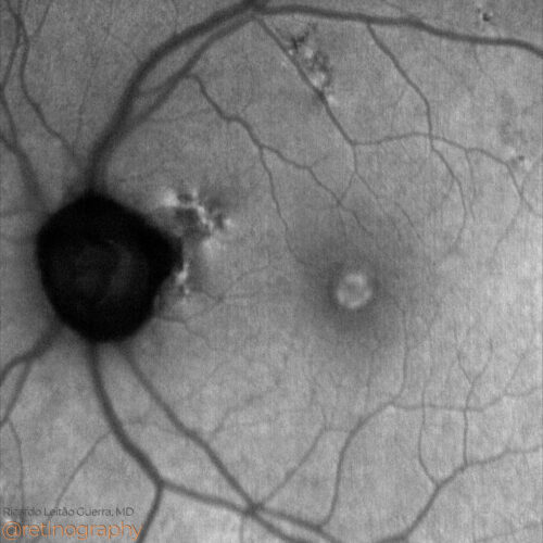 FAF-Green
 True color
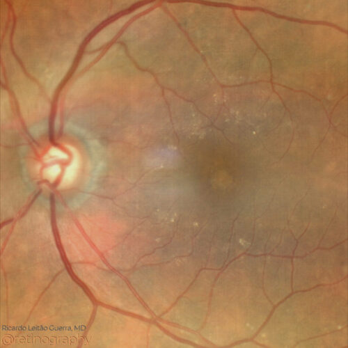 True color - Zoom
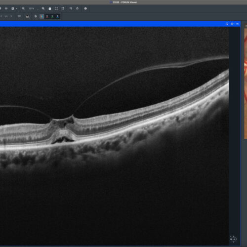 SD-OCT
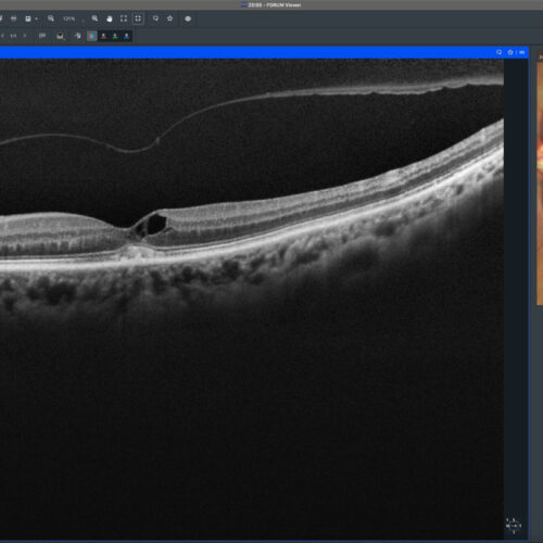 SD-OCT follow up
 Thickness map
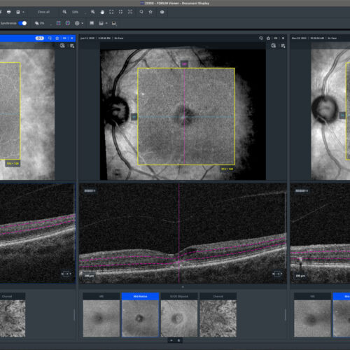 En-face: Mid Retina
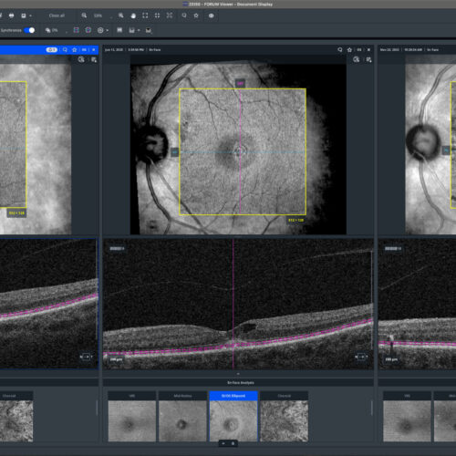 En-face: EZ
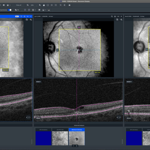 En-face: Minimum intensity
Acquired vitelliform lesions can develop due to vitreomacular traction syndrome. Optical Coherence Tomography (OCT) reveals the presence of subretinal hyperreflective material and vitreomacu...
Acquired vitelliform lesions can develop due to vitreomacular traction syndrome. Optical Coherence Tomography (OCT) reveals the presence of subretinal hyperreflective material and vitreomacu...
Acquired vitelliform lesions can develop due to vitreomacular traction syndrome. Optical Coherence Tomography (OCT) reveals the presence of subretinal hyperreflective material and vitreomacular interface abnormalities. Fundus autofluorescence (FAF) highlights the accumulation of lipofuscin, providing further detail on the lesion’s composition and progression.
#VitelliformLesion #VitreomacularTraction #OCT #FAF #retina #oftalmo #ophthalmology #oftalmologia #oftalmología #ophtalmologie #офтальмологія #офтальмология #οφθαλμολογία #retinography2024 #CIRRUS6000 #CLARUS700 #ZEISSRETINAWORKFLOW
BackRead More -
Acquired vitelliform lesion
Ricardo Leitão Guerra, MD 92yo 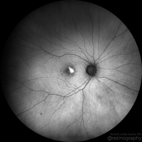 FAF Green
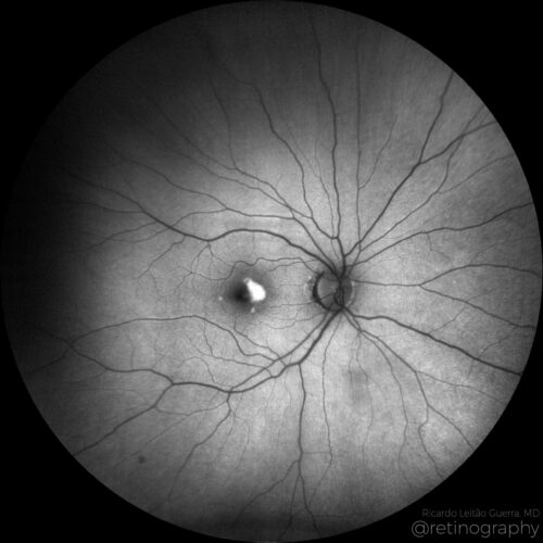 FAF Blue
Acquired vitelliform lesions (AVL) linked to Age-related Macular Degeneration (AMD) present as yellowish round subretinal deposits detectable on fundus examination. Fundus Autofluorescence (...
Acquired vitelliform lesions (AVL) linked to Age-related Macular Degeneration (AMD) present as yellowish round subretinal deposits detectable on fundus examination. Fundus Autofluorescence (...
Acquired vitelliform lesions (AVL) linked to Age-related Macular Degeneration (AMD) present as yellowish round subretinal deposits detectable on fundus examination. Fundus Autofluorescence (FAF) imaging is crucial for AVL detection, showing increased autofluorescence due to lipofuscin accumulation in retinal pigment epithelial cells. These lesions differ from typical AMD deposits and can signify an advanced AMD stage. Understanding AVL’s FAF characteristics helps in differential diagnosis and monitoring AMD progression.
Disclosure: All images featured in this post were acquired and analyzed using devices integrated within the Zeiss Retina Workflow. This ensures high-quality, detailed visual data for comprehensive assessment.
BackRead More