


Tag:
BRAO
-
Branch retinal artery occlusion
Ricardo Leitão Guerra, MD 71yo
71yo  FAF
FAF FA - Early phase
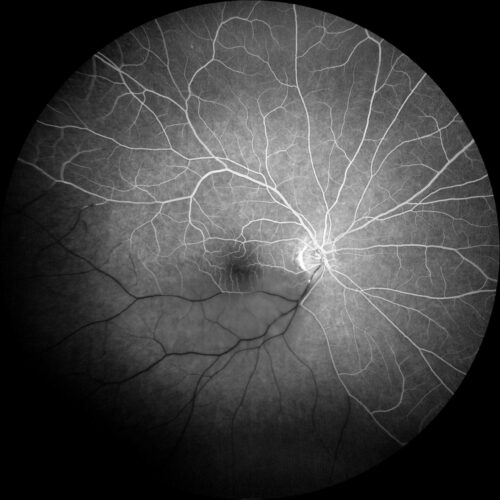 FA - Mid phase
 FA - Late phase
FIGURE 1) True color fundus image allow the identification of a yellowish cholesterol emboli (Hollenhorst plaque) obstructing the inferno-temporal artery branch and the retinal whitening of...
FIGURE 1) True color fundus image allow the identification of a yellowish cholesterol emboli (Hollenhorst plaque) obstructing the inferno-temporal artery branch and the retinal whitening of...
FIGURE 1) True color fundus image allow the identification of a yellowish cholesterol emboli (Hollenhorst plaque) obstructing the inferno-temporal artery branch and the retinal whitening of the affected area. FIGURE 2) Fundus autofluorescence presented a marked hyperautfluorescence of the cholesterol emboli. FIGURE 3, 4 and 5) Fluorescein angiography is extremely helpful determining the extension of the ischemia, as well as the presence of retrograde filling, which is a sign of a better prognosis.
Disclosure: All images featured in this post were acquired and analyzed using devices integrated within the Zeiss Retina Workflow. This ensures high-quality, detailed visual data for comprehensive assessment.
BackRead More