


Tag:
choroidal rupture
-
Choroidal rupture
Ricardo Leitão Guerra, MD 15yo
15yo 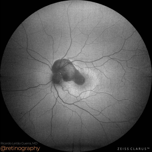 FAF-Green
Choroidal rupture caused by blunt trauma often presents as a crescent-shaped tear in the choroid, Bruch’s membrane, and retinal pigment epithelium (RPE). Subretinal hemorrhage is commonly ...
Choroidal rupture caused by blunt trauma often presents as a crescent-shaped tear in the choroid, Bruch’s membrane, and retinal pigment epithelium (RPE). Subretinal hemorrhage is commonly ...
Choroidal rupture caused by blunt trauma often presents as a crescent-shaped tear in the choroid, Bruch’s membrane, and retinal pigment epithelium (RPE). Subretinal hemorrhage is commonly associated, complicating visualization of the rupture. Optical Coherence Tomography (OCT) and fundus imaging are crucial for identifying the rupture and assessing the extent of subretinal blood. Prompt monitoring is essential to manage complications such as neovascularization.
#ChoroidalRupture #BluntTrauma #SubretinalBlood #OCT #RetinaImaging #TraumaOphthalmology #retina #oftalmo #ophthalmology #oftalmologia #oftalmología #ophtalmologie #офтальмологія #офтальмология #οφθαλμολογία #retinography2024 #CIRRUS6000 #CLARUS700 #ZEISSRETINAWORKFLOW
BackRead More -
AMD – Soft drusen
Ricardo Leitão Guerra, MD 83yo 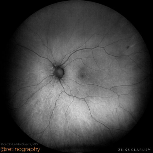 FAF-Green
 NIR & SD-OCT
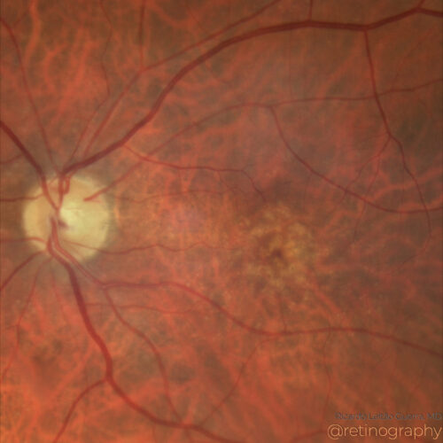 True color - Zoom
 FAF-Green (ZOOM)
Age-related macular degeneration (AMD) with soft drusen involves the accumulation of extracellular material beneath the retina, leading to vision loss. Fundus autofluorescence (FAF) imaging ...
Age-related macular degeneration (AMD) with soft drusen involves the accumulation of extracellular material beneath the retina, leading to vision loss. Fundus autofluorescence (FAF) imaging ...
Age-related macular degeneration (AMD) with soft drusen involves the accumulation of extracellular material beneath the retina, leading to vision loss. Fundus autofluorescence (FAF) imaging detects changes in the retinal pigment epithelium, while optical coherence tomography (OCT) provides detailed cross-sectional images of drusen. These imaging techniques are essential for early diagnosis, monitoring progression, and guiding treatment.
#retina #oftalmo #ophthalmology #oftalmologia #oftalmología #ophtalmologie #офтальмологія #офтальмология #οφθαλμολογία #retinography2024 #CIRRUS6000 #CLARUS700 #ZEISSRETINAWORKFLOW
BackRead More -
Choroidal rupture
Ricardo Leitão Guerra, MD 32yo 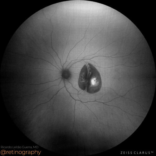 FAF-Green
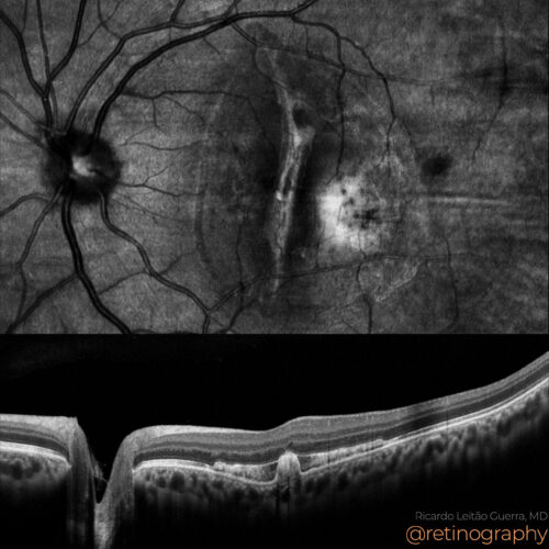 NIR & SD-OCT
Choroidal rupture, often caused by blunt ocular trauma, can lead to subretinal hemorrhage. This condition results in damage to the choroid and underlying structures, causing vision loss. Imm...
Choroidal rupture, often caused by blunt ocular trauma, can lead to subretinal hemorrhage. This condition results in damage to the choroid and underlying structures, causing vision loss. Imm...
Choroidal rupture, often caused by blunt ocular trauma, can lead to subretinal hemorrhage. This condition results in damage to the choroid and underlying structures, causing vision loss. Immediate symptoms may include, blurry vision, and visual field defects. Prompt evaluation and treatment are essential to manage hemorrhage and prevent further complications.
#retina #oftalmo #ophthalmology #oftalmologia #oftalmología #ophtalmologie #офтальмологія #офтальмология #οφθαλμολογία #retinography2024 #CIRRUS6000 #CLARUS700 #ZEISSRETINAWORKFLOW
BackRead More -
Choroidal rupture
Ricardo Leitão Guerra, MD 68yo  FAF - Green
Choroidal rupture is a break in the choroid layer of the eye, commonly resulting from blunt trauma. These ruptures can cause hemorrhages and may lead to complications like choroidal neovascu...
Choroidal rupture is a break in the choroid layer of the eye, commonly resulting from blunt trauma. These ruptures can cause hemorrhages and may lead to complications like choroidal neovascu...
Choroidal rupture is a break in the choroid layer of the eye, commonly resulting from blunt trauma. These ruptures can cause hemorrhages and may lead to complications like choroidal neovascularization. Early detection and management are crucial to prevent vision loss and stabilize ocular health.
Disclosure: All images featured in this post were acquired and analyzed using devices integrated within the Zeiss Retina Workflow. This ensures high-quality, detailed visual data for comprehensive assessment.
BackRead More