


Tag:
CRVO
-
PAMM due to Central Retinal Vein Occlusion
Ricardo Leitão Guerra, MD 63yo
63yo 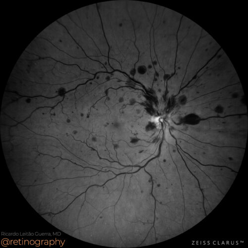 Green channel
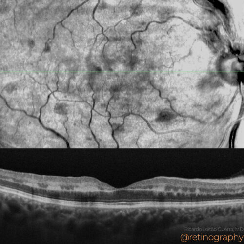 NIR & SD-OCT
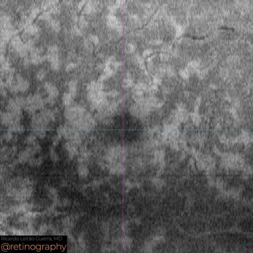 En-face: Mid retina
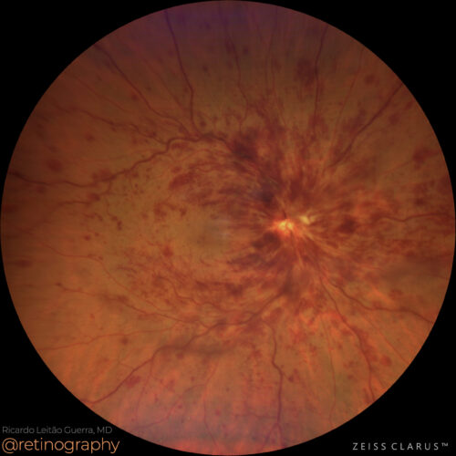 True color
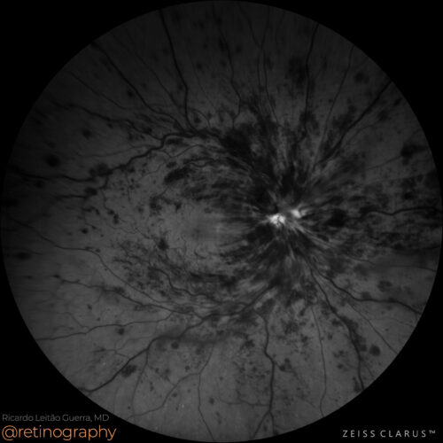 Green Channel
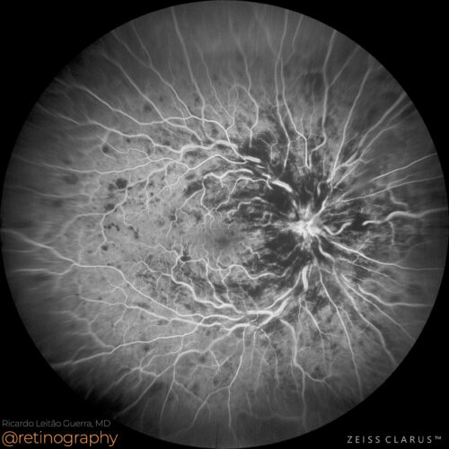 FA: Late phase
Paracentral Acute Middle Maculopathy (PAMM) secondary to Central Retinal Vein Occlusion (CRVO) is characterized by ischemia in the intermediate and deep capillary plexus. En-face OCT reveals...
Paracentral Acute Middle Maculopathy (PAMM) secondary to Central Retinal Vein Occlusion (CRVO) is characterized by ischemia in the intermediate and deep capillary plexus. En-face OCT reveals...
Paracentral Acute Middle Maculopathy (PAMM) secondary to Central Retinal Vein Occlusion (CRVO) is characterized by ischemia in the intermediate and deep capillary plexus. En-face OCT reveals a distinctive “fern-like” pattern, representing hyperreflective bands in the middle retinal layers. This pattern highlights the areas of ischemia, allowing for detailed visualization of retinal damage associated with CRVO.
#PAMM #CRVO #FernLikePattern #EnFaceOCT #RetinaImaging #Ischemia #retina #oftalmo #ophthalmology #oftalmologia #oftalmología #ophtalmologie #офтальмологія #офтальмология #οφθαλμολογία #retinography2024 #CIRRUS6000 #CLARUS700 #ZEISSRETINAWORKFLOW
BackRead More -
Central retinal vein occlusion
Ricardo Leitão Guerra, MD 62yo 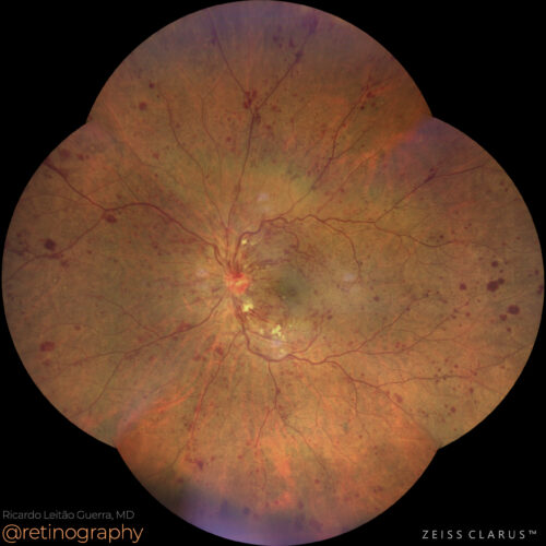 True color: ultra wide-field
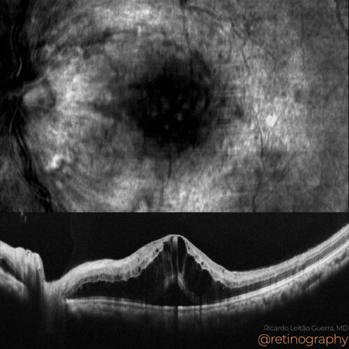 NIR & SD-OCT
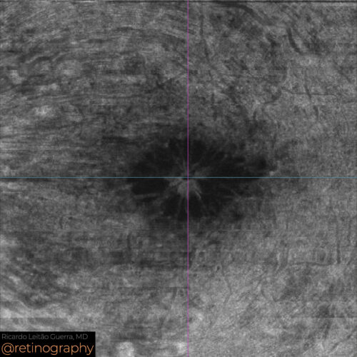 En-face: Mid Retina
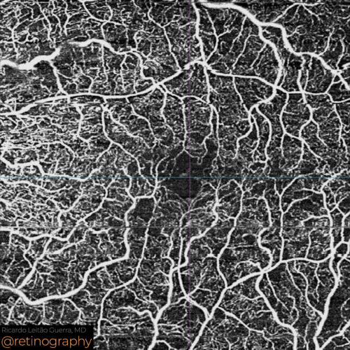 OCT-Angiography
 NIR & SD-OCT
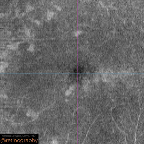 En-face: Mid Retina
Central Retinal Vein Occlusion (CRVO) often leads to macular edema and can be associated with Paracentral Acute Middle Maculopathy (PAMM). PAMM manifests as ischemia in the intermediate reti...
Central Retinal Vein Occlusion (CRVO) often leads to macular edema and can be associated with Paracentral Acute Middle Maculopathy (PAMM). PAMM manifests as ischemia in the intermediate reti...
Central Retinal Vein Occlusion (CRVO) often leads to macular edema and can be associated with Paracentral Acute Middle Maculopathy (PAMM). PAMM manifests as ischemia in the intermediate retinal capillary plexus, visible on OCT as hyperreflective bands. These conditions require careful monitoring and treatment to manage edema and prevent further retinal damage.
#CRVO #MacularEdema #PAMM #RetinaImaging #retina #oftalmo #ophthalmology #oftalmologia #oftalmología #ophtalmologie #офтальмологія #офтальмология #οφθαλμολογία #retinography2024 #CIRRUS6000 #CLARUS700 #ZEISSRETINAWORKFLOW
BackRead More -
Old CRVO
Ricardo Leitão Guerra, MD 72yo 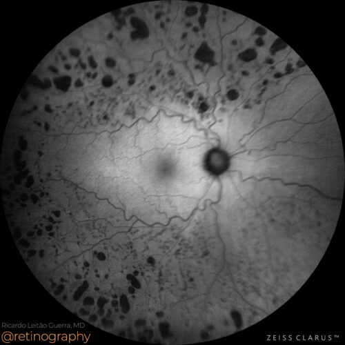 FAF-Green
Old Central Retinal Vein Occlusion (CRVO) can be associated with glaucoma as a significant risk factor due to elevated intraocular pressure contributing to venous occlusion. Panretinal photo...
Old Central Retinal Vein Occlusion (CRVO) can be associated with glaucoma as a significant risk factor due to elevated intraocular pressure contributing to venous occlusion. Panretinal photo...
Old Central Retinal Vein Occlusion (CRVO) can be associated with glaucoma as a significant risk factor due to elevated intraocular pressure contributing to venous occlusion. Panretinal photocoagulation (PRP) is often employed to treat ischemic areas and reduce the risk of neovascular complications, thereby stabilizing the retina and preventing further vision loss.
#CRVO #Glaucoma #RiskFactor #PanretinalPhotocoagulation #PRP #RetinaImaging #retina #oftalmo #ophthalmology #oftalmologia #oftalmología #ophtalmologie #офтальмологія #офтальмология #οφθαλμολογία #retinography2024 #CIRRUS6000 #CLARUS700 #ZEISSRETINAWORKFLOW
BackRead More -
Central retinal vein occlusion
Ricardo Leitão Guerra, MD 36yo 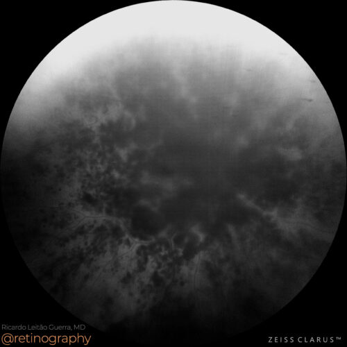 FAF-Green
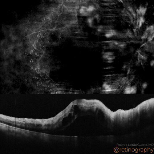 NIR & SD-OCT
Central Retinal Vein Occlusion (CRVO) in a young patient necessitates thorough systemic investigation. Potential underlying causes such as coagulopathies, autoimmune disorders, and systemic ...
Central Retinal Vein Occlusion (CRVO) in a young patient necessitates thorough systemic investigation. Potential underlying causes such as coagulopathies, autoimmune disorders, and systemic ...
Central Retinal Vein Occlusion (CRVO) in a young patient necessitates thorough systemic investigation. Potential underlying causes such as coagulopathies, autoimmune disorders, and systemic hypertension should be explored to identify and manage any contributing factors. Early systemic evaluation is crucial for appropriate treatment and to prevent recurrence or complications.
#CRVO #SystemicInvestigation #YoungPatient #Coagulopathy #Retina #Ophthalmology #RetinaImaging #retina #oftalmo #ophthalmology #oftalmologia #oftalmología #ophtalmologie #офтальмологія #офтальмология #οφθαλμολογία #retinography2024 #CIRRUS6000 #CLARUS700 #ZEISSRETINAWORKFLOW
BackRead More -
Ischemic CRVO
Ricardo Leitão Guerra, MD 63yo
63yo 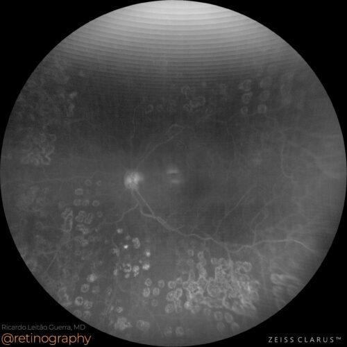 Blue channel
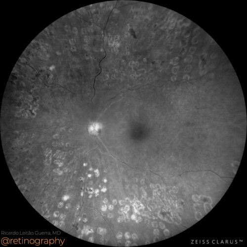 Green channel
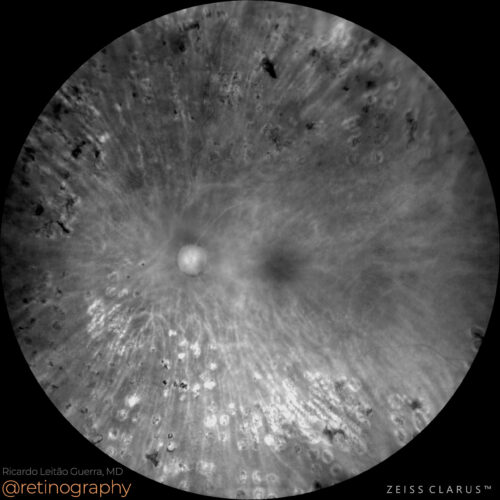 Red channel
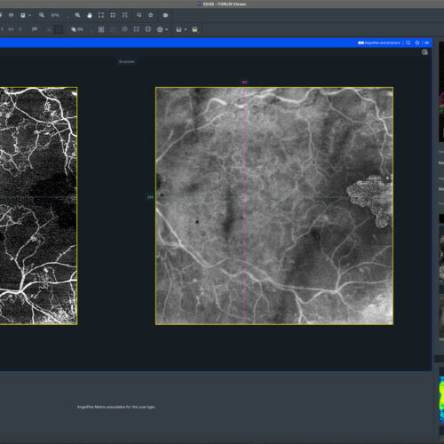 OCT-Angiography
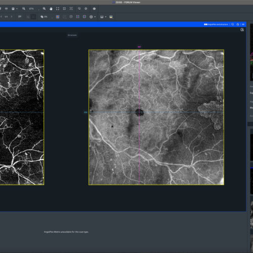 OCT-Angiography
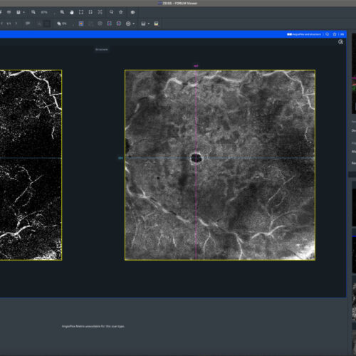 OCT-Angiography
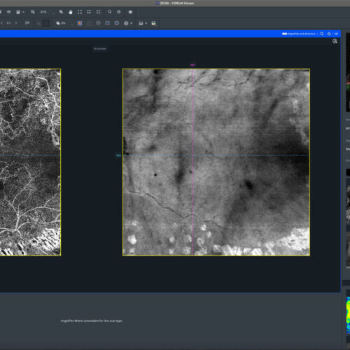 OCT-Angiography
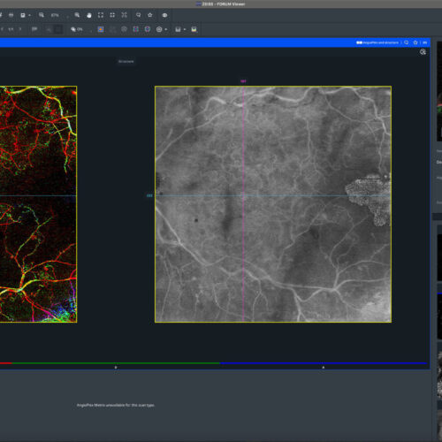 OCT-Angiography
Ischemic Central Retinal Vein Occlusion (CRVO) is a severe form of retinal vein occlusion characterized by extensive retinal ischemia. Wide-field Optical Coherence Tomography Angiography (OC...
Ischemic Central Retinal Vein Occlusion (CRVO) is a severe form of retinal vein occlusion characterized by extensive retinal ischemia. Wide-field Optical Coherence Tomography Angiography (OC...
Ischemic Central Retinal Vein Occlusion (CRVO) is a severe form of retinal vein occlusion characterized by extensive retinal ischemia. Wide-field Optical Coherence Tomography Angiography (OCTA) offers a more extensive view of the retinal vasculature compared to standard OCTA, capturing regions beyond the posterior pole. Although it doesn’t cover the entire retinal periphery, it effectively reveals ischemic areas beyond the central macula, crucial for understanding and managing ischemic conditions like Central Retinal Vein Occlusion (CRVO).
#retina #oftalmo #ophthalmology #oftalmologia #oftalmología #ophtalmologie #офтальмологія #офтальмология #οφθαλμολογία #retinography2024 #CIRRUS6000 #CLARUS700 #ZEISSRETINAWORKFLOW
BackRead More -
Central retinal vein occlusion
Ricardo Leitão Guerra, MD 79yo 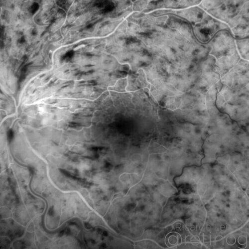 FA: Early phase
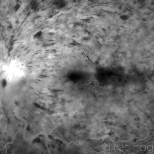 FA: Late phase
Central Retinal Vein Occlusion (CRVO) manifests as retinal hemorrhages and venous engorgement. Fluorescein angiography is vital for assessing retinal perfusion, particularly highlighting are...
Central Retinal Vein Occlusion (CRVO) manifests as retinal hemorrhages and venous engorgement. Fluorescein angiography is vital for assessing retinal perfusion, particularly highlighting are...
Central Retinal Vein Occlusion (CRVO) manifests as retinal hemorrhages and venous engorgement. Fluorescein angiography is vital for assessing retinal perfusion, particularly highlighting areas of non-perfusion like the horizontal raphe in this case. The lack of perfusion indicates ischemia, guiding the prognosis and treatment strategy.
Disclosure: All images featured in this post were acquired and analyzed using devices integrated within the Zeiss Retina Workflow. This ensures high-quality, detailed visual data for comprehensive assessment.
BackRead More