


Tag:
Cystoid macular edema
-
CME in Retinitis pigmentosa
Ricardo Leitão Guerra, MD 32yo
32yo 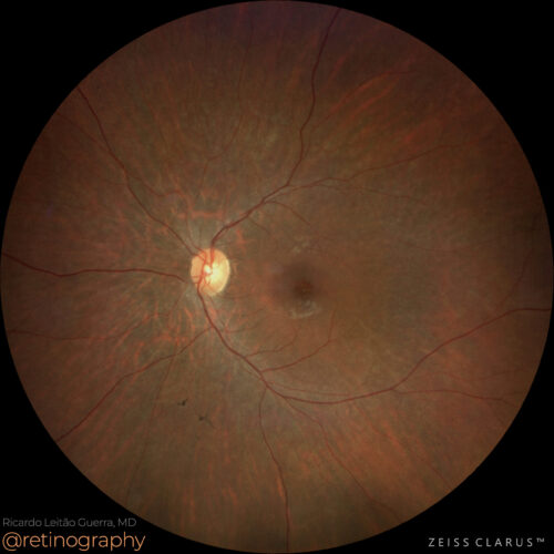 True color
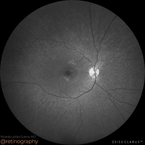 Green channel
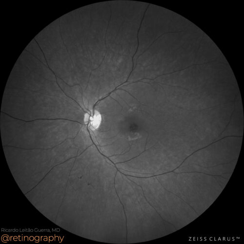 Green channel
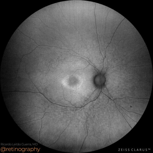 FAF-Green
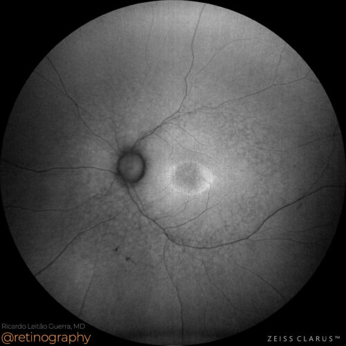 FAF-Green
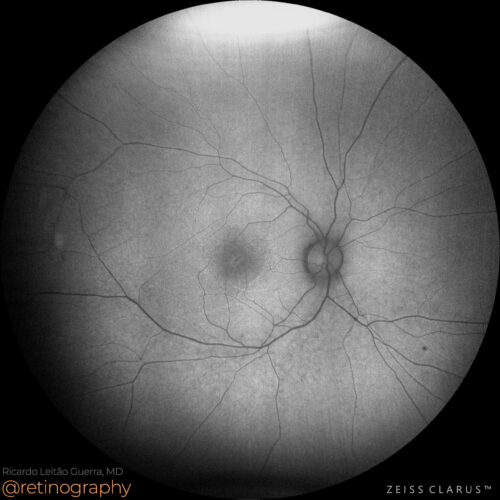 FAF-Blue
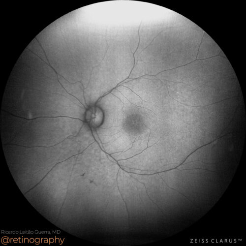 FAF-Blue
In retinitis pigmentosa (RP) with cystoid macular edema (CME), the use of split RGB channels in imaging can be particularly informative. The blue and green channels enhance the visibility of...
In retinitis pigmentosa (RP) with cystoid macular edema (CME), the use of split RGB channels in imaging can be particularly informative. The blue and green channels enhance the visibility of...
In retinitis pigmentosa (RP) with cystoid macular edema (CME), the use of split RGB channels in imaging can be particularly informative. The blue and green channels enhance the visibility of CME, highlighting the fluid-filled cystic spaces in the macula. These channels provide a clearer view of the extent and location of the edema, aiding in diagnosis and management.
#RetinitisPigmentosa #CME #RGBChannels #BlueChannel #GreenChannel #RetinaImaging #retina #oftalmo #ophthalmology #oftalmologia #oftalmología #ophtalmologie #офтальмологія #офтальмология #οφθαλμολογία #retinography2024 #CIRRUS6000 #CLARUS700 #ZEISSRETINAWORKFLOW
BackRead More -
Cystoid macular edema
Ricardo Leitão Guerra, MD 65yo
65yo 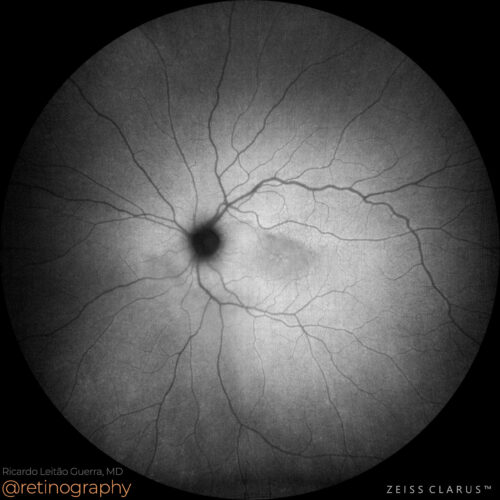 FAF-Green
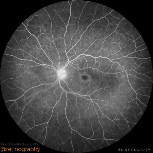 FA: Early phase
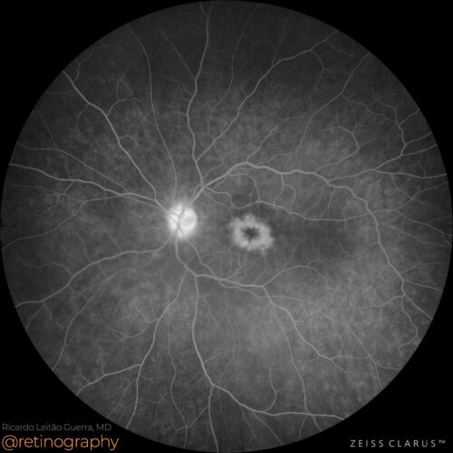 FA: Late phase
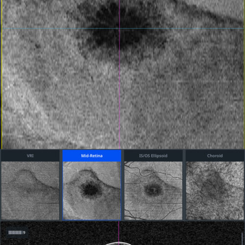 En-face: Mid Retina
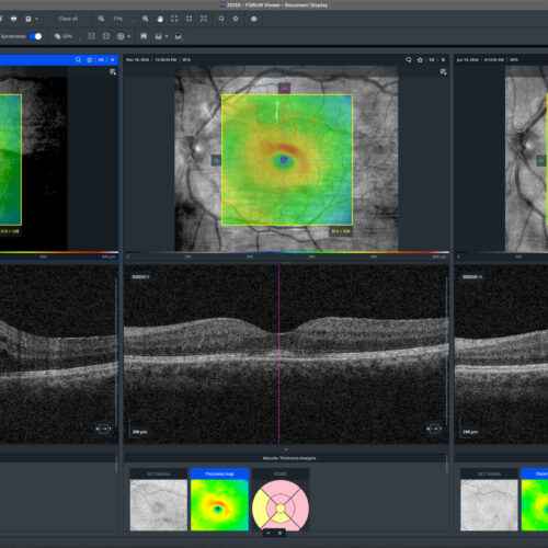 Thickness map: Evolution
Cystoid macular edema (CME) after phacoemulsification can be effectively treated with topical steroids and nonsteroidal anti-inflammatory drugs (NSAIDs). These treatments reduce inflammation...
Cystoid macular edema (CME) after phacoemulsification can be effectively treated with topical steroids and nonsteroidal anti-inflammatory drugs (NSAIDs). These treatments reduce inflammation...
Cystoid macular edema (CME) after phacoemulsification can be effectively treated with topical steroids and nonsteroidal anti-inflammatory drugs (NSAIDs). These treatments reduce inflammation and fluid accumulation in the macula, helping to restore visual acuity and prevent further complications.
#CME #Phacoemulsification #TopicalSteroids #NSAIDs #RetinaImaging #retina #oftalmo #ophthalmology #oftalmologia #oftalmología #ophtalmologie #офтальмологія #офтальмология #οφθαλμολογία #retinography2024 #CIRRUS6000 #CLARUS700 #ZEISSRETINAWORKFLOW
BackRead More -
CME and Neovascular AMD
Ricardo Leitão Guerra, MD 71yo 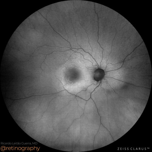 FAF-Green
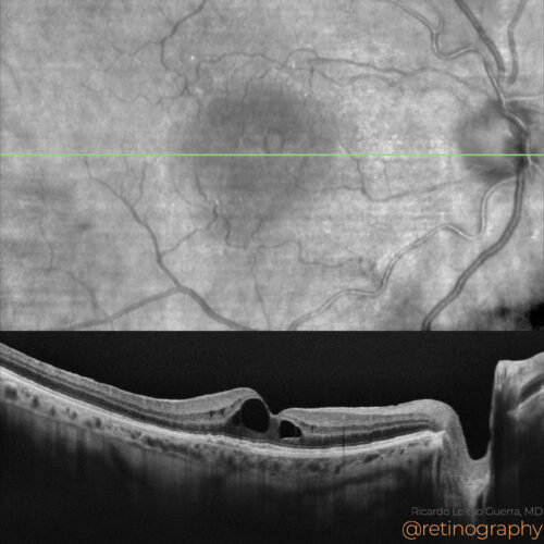 NIR & SD-OCT
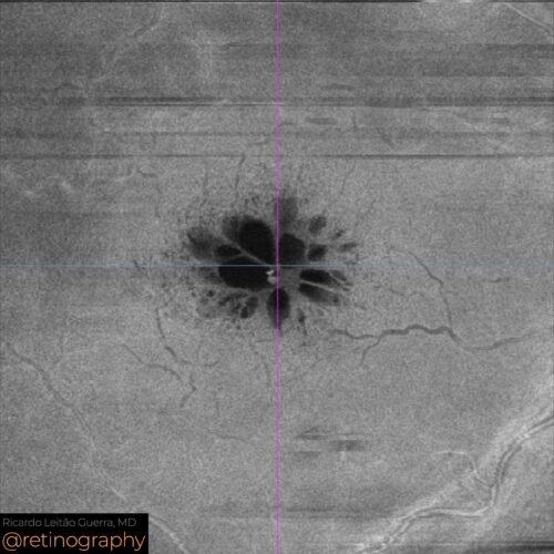 En-face: Mid Retina
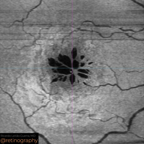 En-face: Minimum intensity
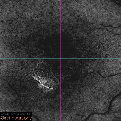 OCT-Angiography
Cystoid macular edema (CME) following phacoemulsification in a patient with neovascular age-related macular degeneration (AMD) and Type 1 neovascular membrane requires careful management. Op...
Cystoid macular edema (CME) following phacoemulsification in a patient with neovascular age-related macular degeneration (AMD) and Type 1 neovascular membrane requires careful management. Op...
Cystoid macular edema (CME) following phacoemulsification in a patient with neovascular age-related macular degeneration (AMD) and Type 1 neovascular membrane requires careful management. Optical Coherence Tomography (OCT) can be used to monitor the edema and the neovascular membrane. Treatment may include anti-VEGF injections and corticosteroids to reduce inflammation and manage the edema.
#CME #NeovascularAMD #Type1NeovascularMembrane #Phacoemulsification #OCT #AntiVEGF #RetinaImaging #retina #oftalmo #ophthalmology #oftalmologia #oftalmología #ophtalmologie #офтальмологія #офтальмология #οφθαλμολογία #retinography2024 #CIRRUS6000 #CLARUS700 #ZEISSRETINAWORKFLOW
BackRead More -
Cystoid Macular Edema
Ricardo Leitão Guerra, MD 74yo 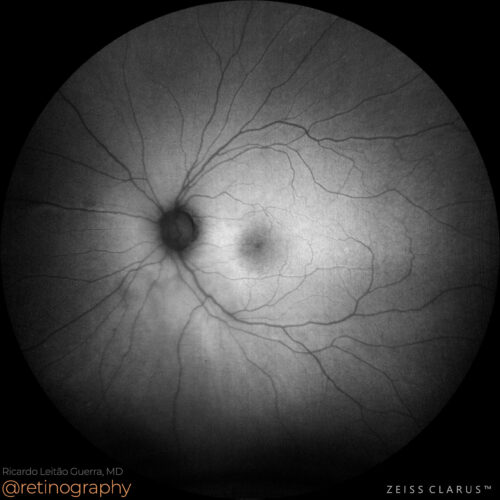 FAF-Green
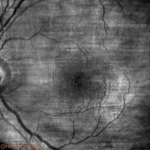 NIR
NIR FA: Mid phase
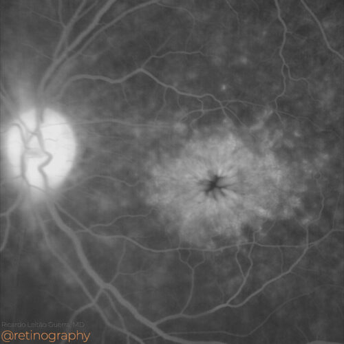 FA: Late phase
Cystoid macular edema (CME) is a common complication following cataract surgery, also known as pseudophakic macular edema. This condition results from inflammation and fluid accumulation in ...
Cystoid macular edema (CME) is a common complication following cataract surgery, also known as pseudophakic macular edema. This condition results from inflammation and fluid accumulation in ...
Cystoid macular edema (CME) is a common complication following cataract surgery, also known as pseudophakic macular edema. This condition results from inflammation and fluid accumulation in the macula, leading to visual impairment. Treatment typically involves topical nonsteroidal anti-inflammatory drugs (NSAIDs) and corticosteroids to reduce inflammation and restore visual function.
#CME #CataractSurgery #PseudophakicMacularEdema #TopicalTherapy #NSAIDs #Corticosteroids #RetinaImaging #retina #oftalmo #ophthalmology #oftalmologia #oftalmología #ophtalmologie #офтальмологія #офтальмология #οφθαλμολογία #retinography2024 #CIRRUS6000 #CLARUS700 #ZEISSRETINAWORKFLOW
BackRead More -
Cystoid Macular Edema
Ricardo Leitão Guerra, MD 74yo 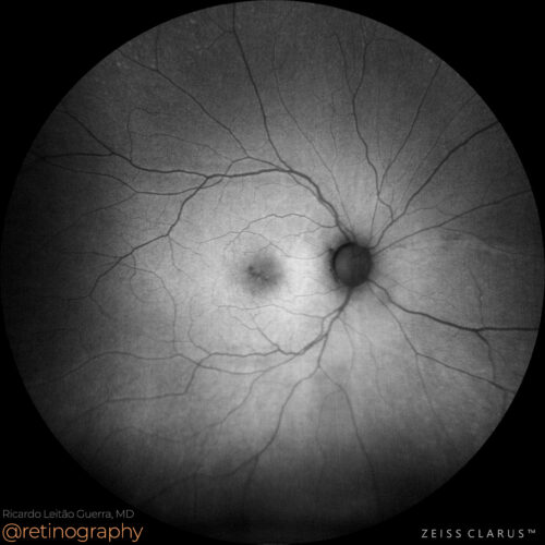 FAF-Green
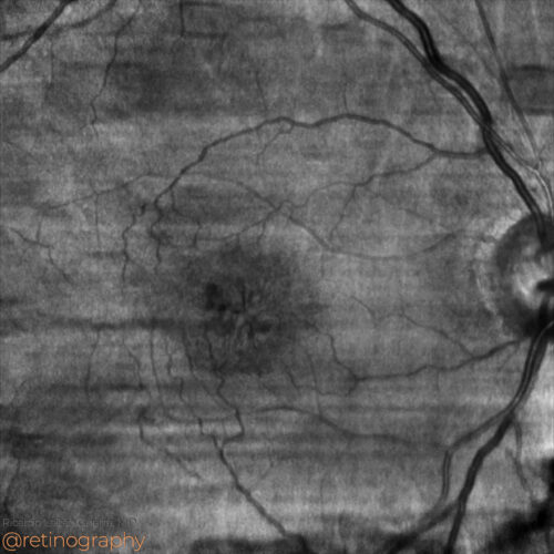 NIR
NIR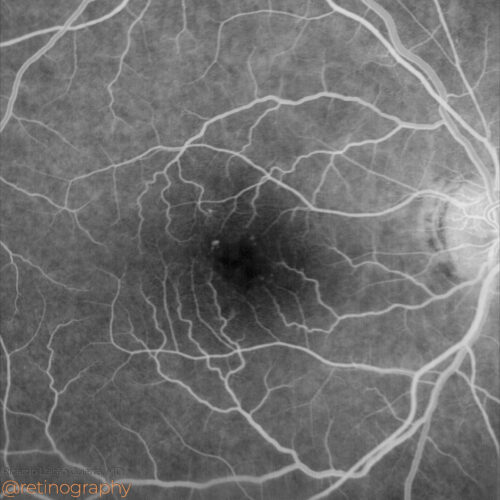 FA: Early phase
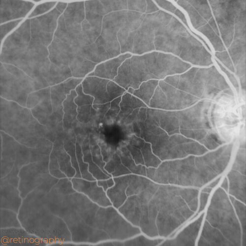 FA: Mid phase
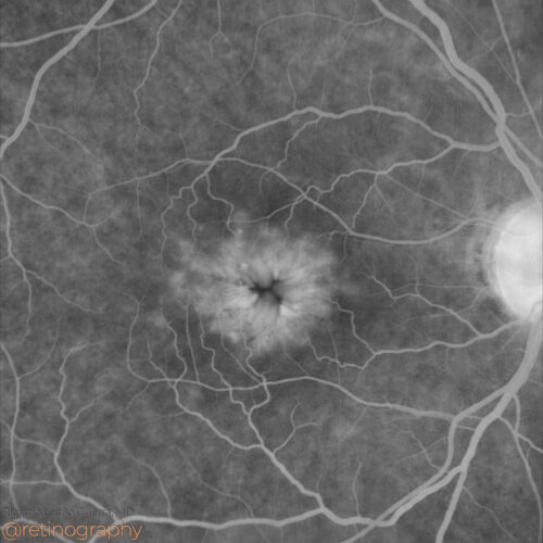 FA: Late phase
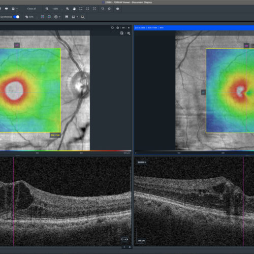 Thickness map: Follow up
Cystoid macular edema (CME) is characterized by fluid-filled cystic spaces within the macula. Fluorescein angiography typically reveals a petalloid pattern of dye leakage in the macula. Topi...
Cystoid macular edema (CME) is characterized by fluid-filled cystic spaces within the macula. Fluorescein angiography typically reveals a petalloid pattern of dye leakage in the macula. Topi...
Cystoid macular edema (CME) is characterized by fluid-filled cystic spaces within the macula. Fluorescein angiography typically reveals a petalloid pattern of dye leakage in the macula. Topical therapy, including nonsteroidal anti-inflammatory drugs (NSAIDs) or corticosteroids, can be effective in reducing inflammation and fluid accumulation, thereby improving visual acuity.
#CME #FluoresceinAngiography #PetalloidPattern #TopicalTherapy #NSAIDs #Corticosteroids #RetinaImaging #retina #oftalmo #ophthalmology #oftalmologia #oftalmología #ophtalmologie #офтальмологія #офтальмология #οφθαλμολογία #retinography2024 #CIRRUS6000 #CLARUS700 #ZEISSRETINAWORKFLOW
BackRead More -
Cystoid macular edema
Ricardo Leitão Guerra, MD 75yo 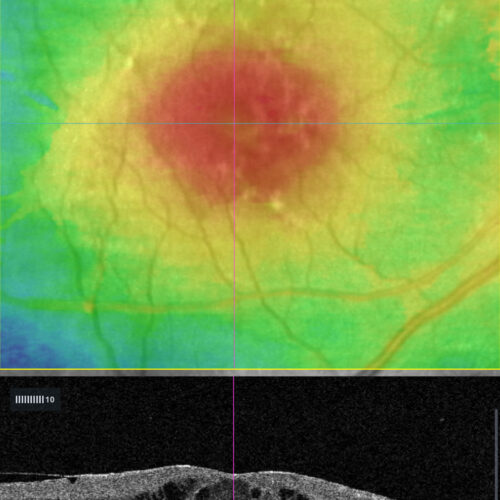 OCT: Thickness map
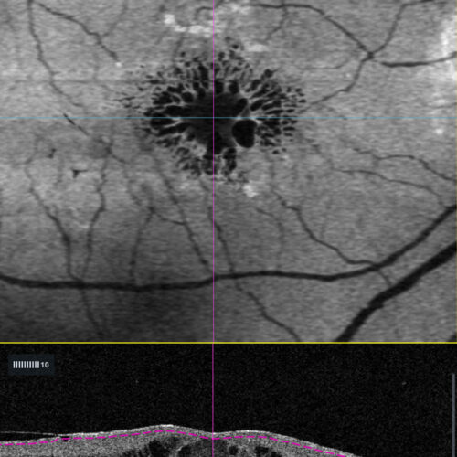 OCT En-face: Minimun intensity
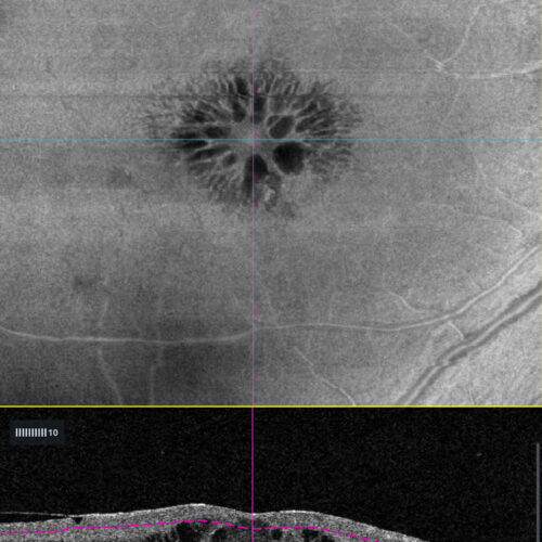 OCT En-face: Mid-retina
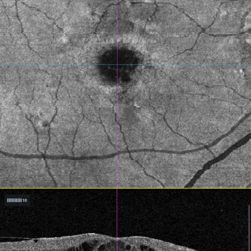 OCT En-face: EZ
Cystoid macular edema (CME) is characterized by fluid-filled cystic spaces in the macula. Enface OCT imaging reveals these cystic spaces as hyporeflective areas within the retinal layers, of...
Cystoid macular edema (CME) is characterized by fluid-filled cystic spaces in the macula. Enface OCT imaging reveals these cystic spaces as hyporeflective areas within the retinal layers, of...
Cystoid macular edema (CME) is characterized by fluid-filled cystic spaces in the macula. Enface OCT imaging reveals these cystic spaces as hyporeflective areas within the retinal layers, offering a detailed topographical view of the extent and distribution of edema. This imaging technique aids in assessing treatment response and disease progression.
#retina #oftalmo #ophthalmology #oftalmologia #oftalmología #ophtalmologie #офтальмологія #офтальмология #οφθαλμολογία #retinography2024 #CIRRUS6000 #CLARUS700 #ZEISSRETINAWORKFLOW
BackRead More