


Tag:
diabetic macular edema
-
Diabetic macular edema
Ricardo Leitão Guerra, MD 79yo
79yo 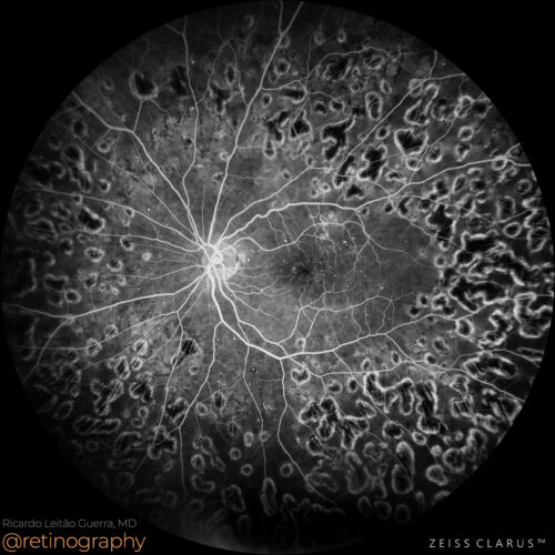 FA: Early phase
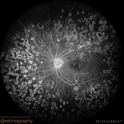 FA: Late phase
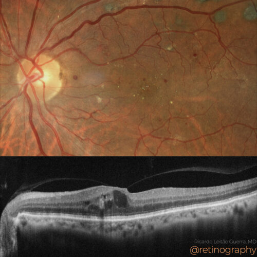 True color & SD-OCT
Diabetic macular edema (DME) is characterized by retinal thickening and fluid accumulation in the macula, often leading to vision loss. Fluorescein angiography (FA) is crucial for evaluating...
Diabetic macular edema (DME) is characterized by retinal thickening and fluid accumulation in the macula, often leading to vision loss. Fluorescein angiography (FA) is crucial for evaluating...
Diabetic macular edema (DME) is characterized by retinal thickening and fluid accumulation in the macula, often leading to vision loss. Fluorescein angiography (FA) is crucial for evaluating DME, as it reveals areas of capillary leakage, microaneurysms, and zones of ischemia. This imaging helps to assess the extent of vascular damage, guiding treatment with options like anti-VEGF injections and laser therapy.
#DiabeticMacularEdema #DME #FluoresceinAngiography #FA #RetinaImaging #retina #oftalmo #ophthalmology #oftalmologia #oftalmología #ophtalmologie #офтальмологія #офтальмология #οφθαλμολογία #retinography2024 #CIRRUS6000 #CLARUS700 #ZEISSRETINAWORKFLOW
BackRead More -
Diabetic Macular Edema
Ricardo Leitão Guerra, MD 37yo
37yo  FAF-Green
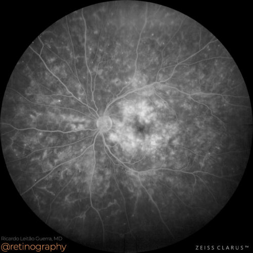 FA: Late phase
Diabetic macular edema (DME) with longstanding subretinal fluid (SRF) can lead to hyperautofluorescence on fundus autofluorescence (FAF) imaging. This hyperautofluorescence indicates chronic...
Diabetic macular edema (DME) with longstanding subretinal fluid (SRF) can lead to hyperautofluorescence on fundus autofluorescence (FAF) imaging. This hyperautofluorescence indicates chronic...
Diabetic macular edema (DME) with longstanding subretinal fluid (SRF) can lead to hyperautofluorescence on fundus autofluorescence (FAF) imaging. This hyperautofluorescence indicates chronic damage and accumulation of lipofuscin in the retinal pigment epithelium (RPE). It is a potential biomarker of worse prognosis, as it suggests persistent retinal stress and damage.
#DME #SubretinalFluid #Hyperautofluorescence #FAF #Biomarker #WorsePrognosis #RetinaImaging #retina #oftalmo #ophthalmology #oftalmologia #oftalmología #ophtalmologie #офтальмологія #офтальмология #οφθαλμολογία #retinography2024 #CIRRUS6000 #CLARUS700 #ZEISSRETINAWORKFLOW
BackRead More -
Diabetic macular edema
Ricardo Leitão Guerra, MD 37yo 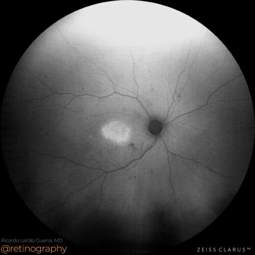 FAF-Green
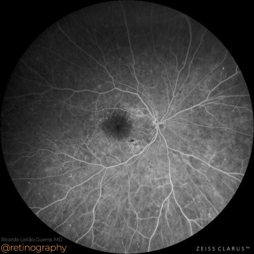 FA: Early phase
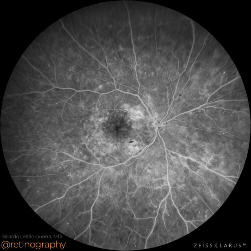 FA: Mid phase
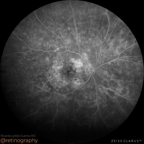 FA: Late phase
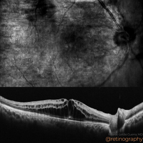 NIR & SD-OCT
Diabetic retinopathy is characterized by increased vascular permeability, leading to leakage of blood and fluid into the retina. Fluorescein angiography (FA) is used to assess this permeabil...
Diabetic retinopathy is characterized by increased vascular permeability, leading to leakage of blood and fluid into the retina. Fluorescein angiography (FA) is used to assess this permeabil...
Diabetic retinopathy is characterized by increased vascular permeability, leading to leakage of blood and fluid into the retina. Fluorescein angiography (FA) is used to assess this permeability, revealing areas of leakage, microaneurysms, and also neovascularization. FA helps in evaluating the extent of retinal damage and guiding treatment decisions.
#DiabeticRetinopathy #VascularPermeability #FluoresceinAngiography #FA #RetinaImaging #retina #oftalmo #ophthalmology #oftalmologia #oftalmología #ophtalmologie #офтальмологія #офтальмология #οφθαλμολογία #retinography2024 #CIRRUS6000 #CLARUS700
BackRead More -
Persistent diabetic macular edema
Ricardo Leitão Guerra, MD 75yo 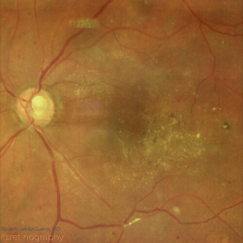 True color: Follow up
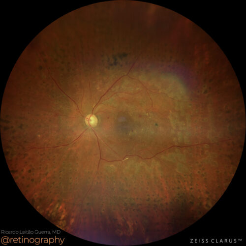 True color - Zoom
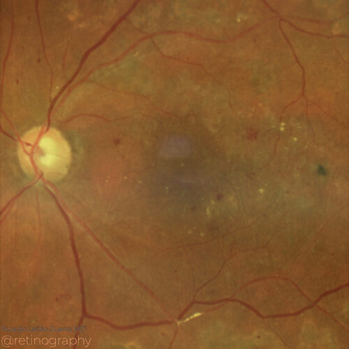 True color - Zoom
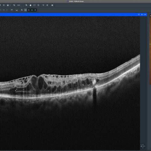 SD-OCT
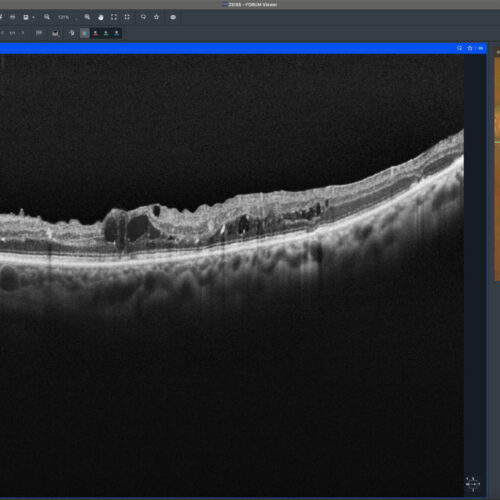 SD-OCT follow up
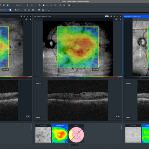 Evolution: Thickness map
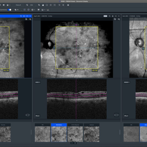 Evolution: En-face
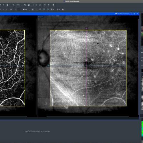 OCT-Angiography
Persistent diabetic macular edema (DME) despite intravitreal anti-VEGF and corticosteroids can be due to traction caused by an epiretinal membrane (ERM). Surgical intervention, such as vitre...
Persistent diabetic macular edema (DME) despite intravitreal anti-VEGF and corticosteroids can be due to traction caused by an epiretinal membrane (ERM). Surgical intervention, such as vitre...
BackRead More -
DME: Telangiectatic capillaries
Ricardo Leitão Guerra, MD 75yo 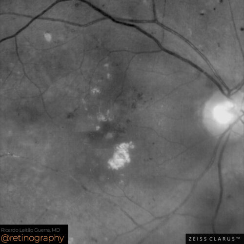 Green channel: Zoom
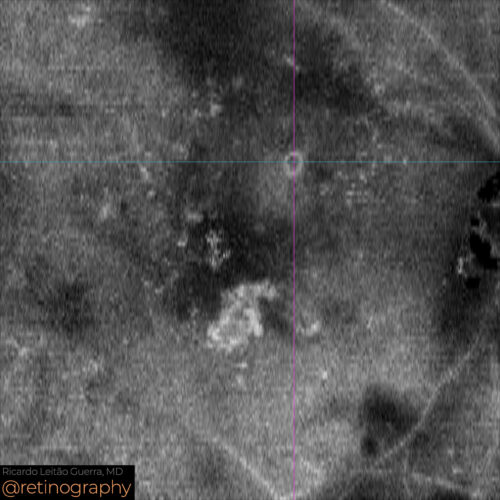 En-face: Mid Retina
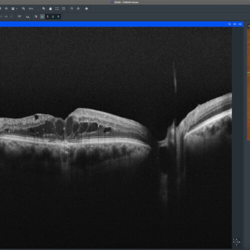 SD-OCT: Baseline
 SD-OCT: Follow up
 SD-OCT: Follow up
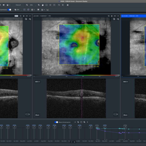 Evolution: Thickness Map
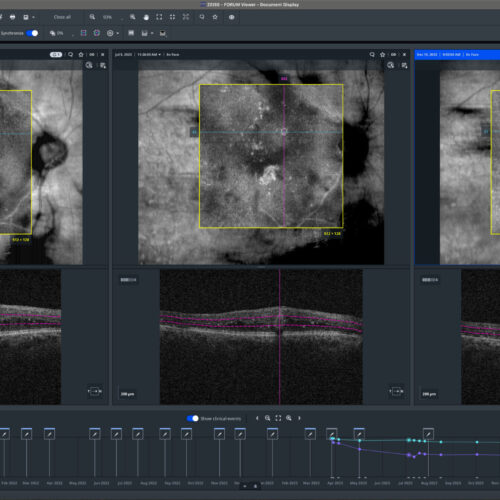 Evolution: En-face
Telangiectatic capillaries (TelCap) can contribute to persistent diabetic macular edema (DME). En-face imaging helps identify these abnormal capillaries, highlighting their distribution and ...
Telangiectatic capillaries (TelCap) can contribute to persistent diabetic macular edema (DME). En-face imaging helps identify these abnormal capillaries, highlighting their distribution and ...
Telangiectatic capillaries (TelCap) can contribute to persistent diabetic macular edema (DME). En-face imaging helps identify these abnormal capillaries, highlighting their distribution and extent. Focal laser photocoagulation is used to target and seal these TelCaps, reducing edema and improving retinal health.
#TelCap #DME #EnFaceImaging #LaserPhotocoagulation #retina #oftalmo #ophthalmology #oftalmologia #oftalmología #ophtalmologie #офтальмологія #офтальмология #οφθαλμολογία #retinography2024 #CIRRUS6000 #CLARUS700 #ZEISSRETINAWORKFLOW
BackRead More -
Diabetic retinopathy: ERM
Ricardo Leitão Guerra, MD 75yo  SD-OCT
 True color
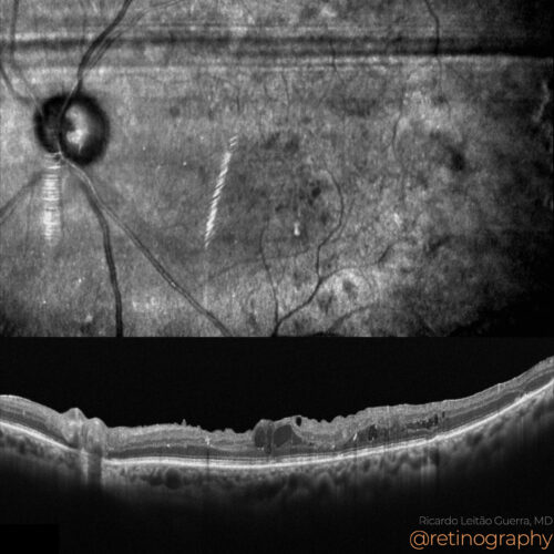 NIR & SD-OCT
 SD-OCT follow up
Diabetic macular edema (DME) refractory to intravitreal treatment, sometimes exacerbated by the presence of an epiretinal membrane (ERM), can significantly impair visual acuity. Standard tre...
Diabetic macular edema (DME) refractory to intravitreal treatment, sometimes exacerbated by the presence of an epiretinal membrane (ERM), can significantly impair visual acuity. Standard tre...
Diabetic macular edema (DME) refractory to intravitreal treatment, sometimes exacerbated by the presence of an epiretinal membrane (ERM), can significantly impair visual acuity. Standard treatments, such as anti-VEGF or corticosteroids, may be insufficient. However, vitrectomy with ERM peeling has been shown to improve visual and anatomical outcomes in these cases by relieving traction on the macula and enhancing fluid absorption.
#retina #oftalmo #ophthalmology #oftalmologia #oftalmología #ophtalmologie #офтальмологія #офтальмология #οφθαλμολογία #retinography2024 #CIRRUS6000 #CLARUS700 #ZEISSRETINAWORKFLOW
BackRead More