


Tag:
macular neovascularization
-
MNV: Best’s vitelliform dystrophy
Ricardo Leitão Guerra, MD 12yo
12yo 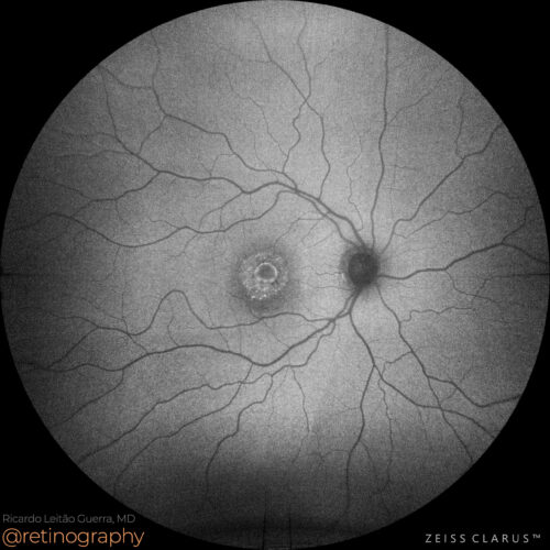 FAF-Green
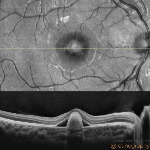 NIR & SD-OCT
In Best’s vitelliform dystrophy, macular neovascularization (MNV) can develop as a complication. Optical Coherence Tomography (OCT) helps detect subretinal or intraretinal fluid and ne...
In Best’s vitelliform dystrophy, macular neovascularization (MNV) can develop as a complication. Optical Coherence Tomography (OCT) helps detect subretinal or intraretinal fluid and ne...
BackRead More -
Neovascular AMD
Ricardo Leitão Guerra, MD 94yo
94yo 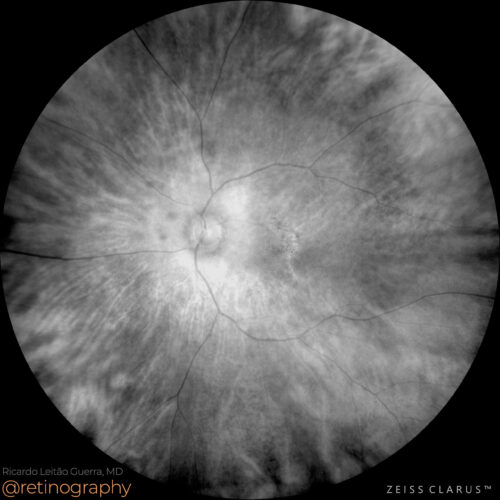 Red channel
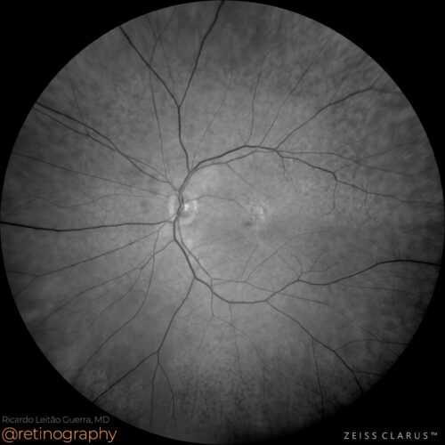 Green channel
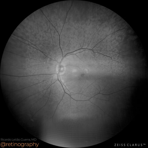 Blue channel
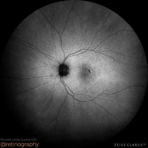 FAF-Green
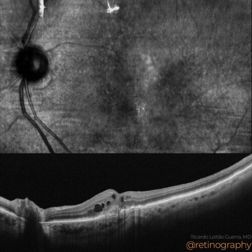 NIR & SD-OCT
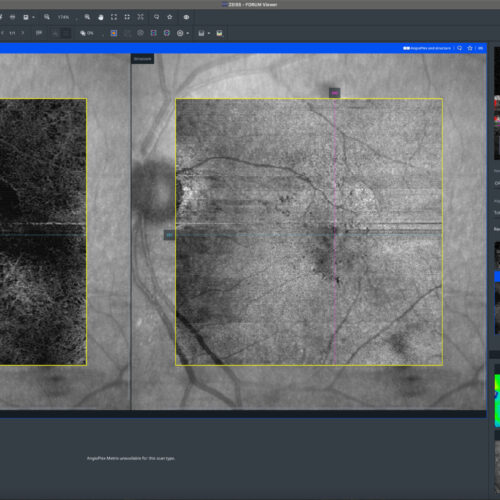 OCT-Angiography
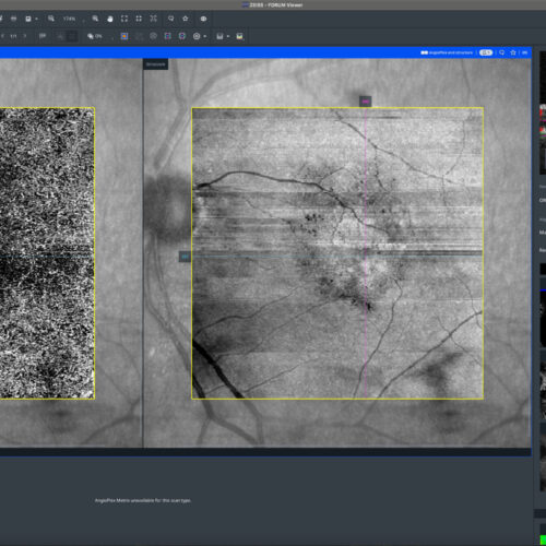 OCT-Angiography
Neovascular age-related macular degeneration (AMD) often presents with pseudodrusen, which can be highlighted using the blue channel in color fundus photography. Optical Coherence Tomography...
Neovascular age-related macular degeneration (AMD) often presents with pseudodrusen, which can be highlighted using the blue channel in color fundus photography. Optical Coherence Tomography...
BackRead More -
Quiescent type 1 MNV
Ricardo Leitão Guerra, MD 80yo 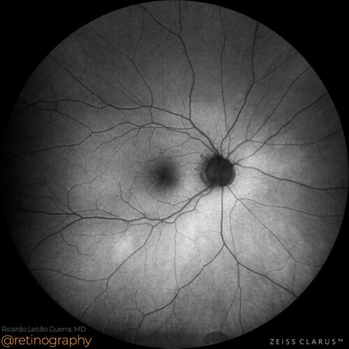 FAF-Green
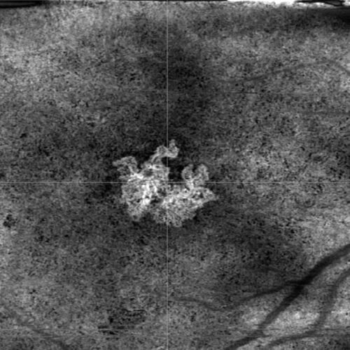 HD 6mm OCT Angiography - Custom sub-RPE segmentation
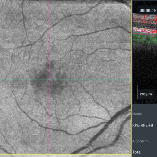 OCT Angiography - Custom sub-RPE segmentation
Quiescent type 1 macular neovascularization (MNV) is a form of neovascular AMD without exudation or hemorrhage. Detection using OCT Angiography (OCTA) involves identifying abnormal blood ves...
Quiescent type 1 macular neovascularization (MNV) is a form of neovascular AMD without exudation or hemorrhage. Detection using OCT Angiography (OCTA) involves identifying abnormal blood ves...
Quiescent type 1 macular neovascularization (MNV) is a form of neovascular AMD without exudation or hemorrhage. Detection using OCT Angiography (OCTA) involves identifying abnormal blood vessels in the sub-RPE space without fluid accumulation. OCTA allows for non-invasive visualization and monitoring, crucial for timely intervention before the onset of exudative changes.
#retina #oftalmo #ophthalmology #oftalmologia #oftalmología #ophtalmologie #офтальмологія #офтальмология #οφθαλμολογία #retinography2024 #CIRRUS6000 #CLARUS700 #ZEISSRETINAWORKFLOW
BackRead More -
Myopic MNV
Ricardo Leitão Guerra, MD 39yo  FAF-Green
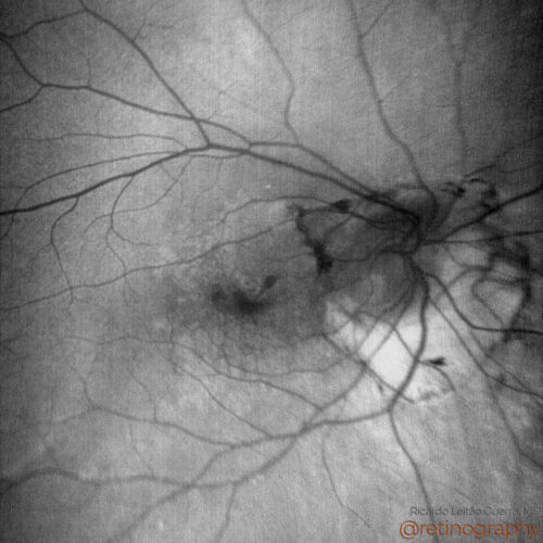 FAF-Blue
 FA: Early phase
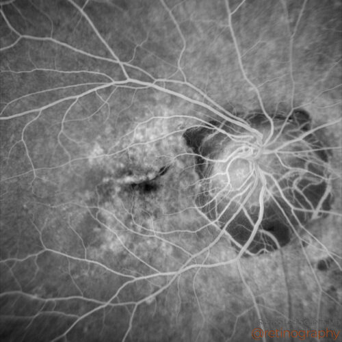 FA: mid phase
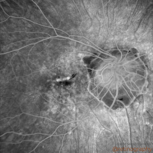 FA: late phase
 FA: late phase
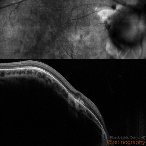 NIR & SD-OCT
Myopic neovascularization, linked to high myopia, shows distinctive features across multiple imaging modalities. OCT reveals subretinal neovascular membranes with fluid. Fluorescein angiogra...
Myopic neovascularization, linked to high myopia, shows distinctive features across multiple imaging modalities. OCT reveals subretinal neovascular membranes with fluid. Fluorescein angiogra...
Myopic neovascularization, linked to high myopia, shows distinctive features across multiple imaging modalities. OCT reveals subretinal neovascular membranes with fluid. Fluorescein angiography highlights leakage from these membranes, while OCT angiography provides detailed vascular imaging without dye use, critical for precise treatment planning.
Disclosure: All images featured in this post were acquired and analyzed using devices integrated within the Zeiss Retina Workflow. This ensures high-quality, detailed visual data for comprehensive assessment.
BackRead More