


Tag:
Multimodal retinal analysis
-
Choroidal nevus
Ricardo Leitão Guerra, MD 51yo
51yo 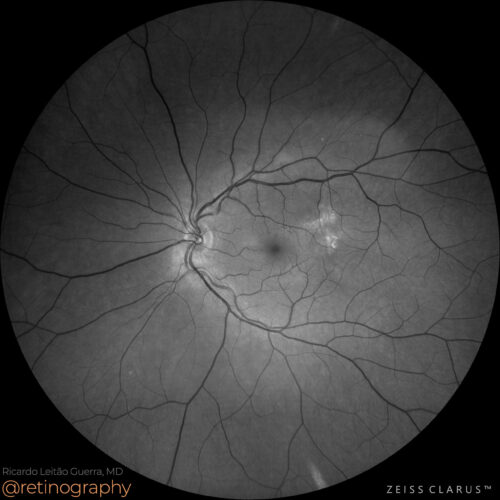 Green channel
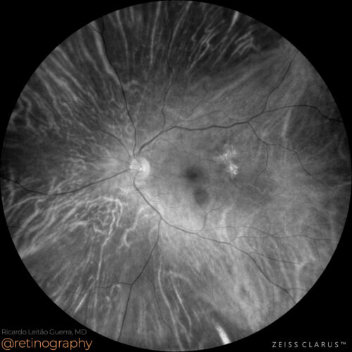 Red channel
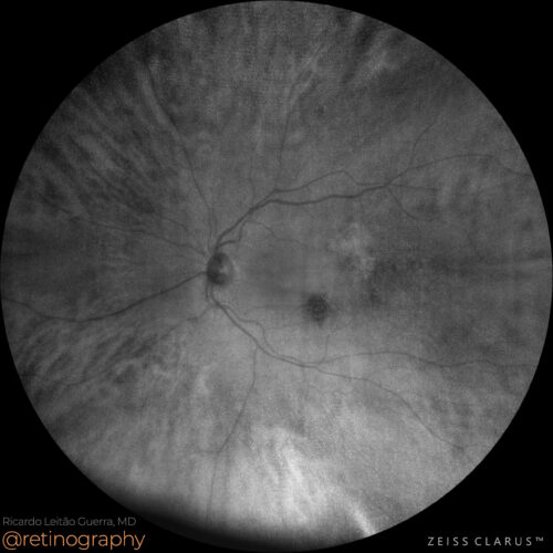 NIR
NIR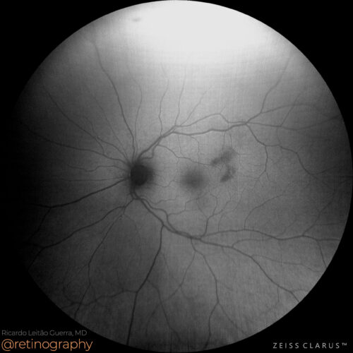 FAF-Green
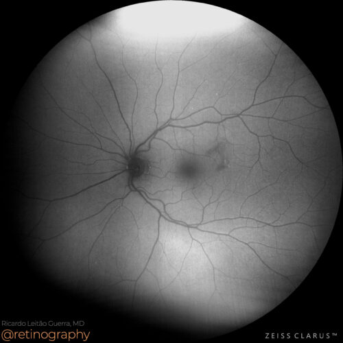 FAF-Blue
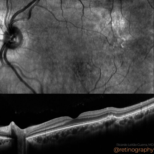 MODELO SITE DIESSY.008
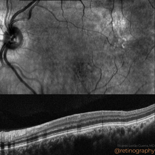 NIR & SD-OCT
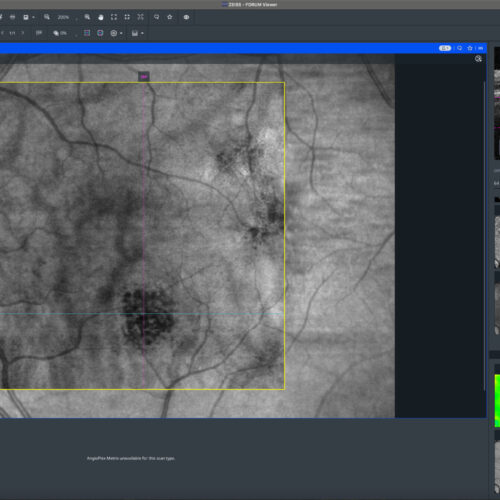 En-face: Choroid
En-face: Choroid
En-face: ChoroidChoroidal nevus is a benign pigmented lesion in the choroid, often monitored for potential malignant transformation. Near-Infrared Reflectance (NIR) imaging and the red channel in color fund...
Choroidal nevus is a benign pigmented lesion in the choroid, often monitored for potential malignant transformation. Near-Infrared Reflectance (NIR) imaging and the red channel in color fund...
Choroidal nevus is a benign pigmented lesion in the choroid, often monitored for potential malignant transformation. Near-Infrared Reflectance (NIR) imaging and the red channel in color fundus photography enhance the visibility of the nevus. Optical Coherence Tomography (OCT) provides detailed cross-sectional images to assess the nevus’s impact on retinal layers. OCT Angiography (OCTA) evaluates the blood flow around the lesion, aiding in detecting any neovascular activity. In this case there is also a quiescent type 1 macular neovascularization at the temporal-superior macular area.
#retina #oftalmo #ophthalmology #oftalmologia #oftalmología #ophtalmologie #офтальмологія #офтальмология #οφθαλμολογία #retinography2024 #CIRRUS6000 #CLARUS700 #ZEISSRETINAWORKFLOW
BackRead More -
Cystoid macular edema
Ricardo Leitão Guerra, MD 75yo 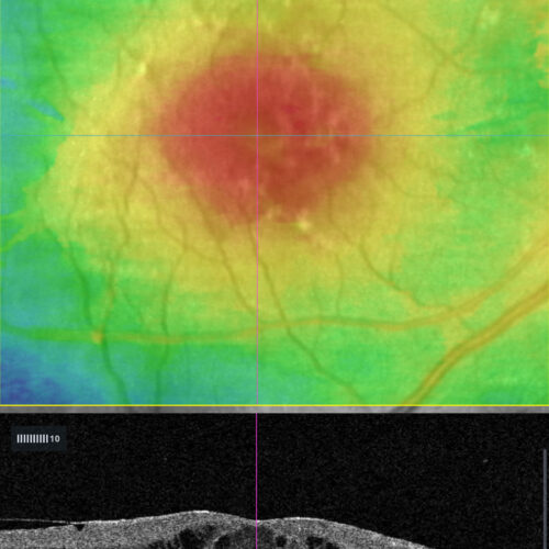 OCT: Thickness map
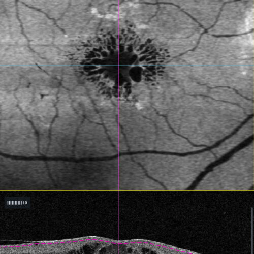 OCT En-face: Minimun intensity
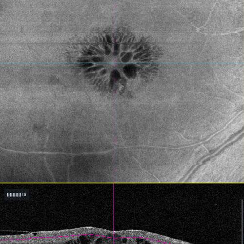 OCT En-face: Mid-retina
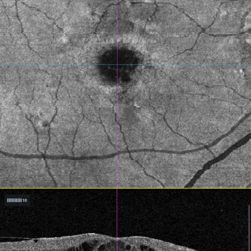 OCT En-face: EZ
Cystoid macular edema (CME) is characterized by fluid-filled cystic spaces in the macula. Enface OCT imaging reveals these cystic spaces as hyporeflective areas within the retinal layers, of...
Cystoid macular edema (CME) is characterized by fluid-filled cystic spaces in the macula. Enface OCT imaging reveals these cystic spaces as hyporeflective areas within the retinal layers, of...
Cystoid macular edema (CME) is characterized by fluid-filled cystic spaces in the macula. Enface OCT imaging reveals these cystic spaces as hyporeflective areas within the retinal layers, offering a detailed topographical view of the extent and distribution of edema. This imaging technique aids in assessing treatment response and disease progression.
#retina #oftalmo #ophthalmology #oftalmologia #oftalmología #ophtalmologie #офтальмологія #офтальмология #οφθαλμολογία #retinography2024 #CIRRUS6000 #CLARUS700 #ZEISSRETINAWORKFLOW
BackRead More -
Quiescent type 1 MNV
Ricardo Leitão Guerra, MD 80yo 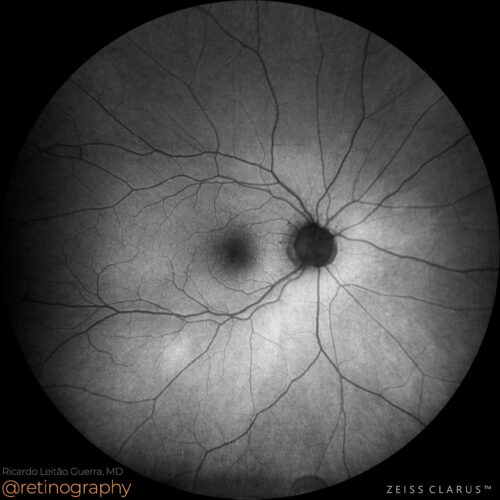 FAF-Green
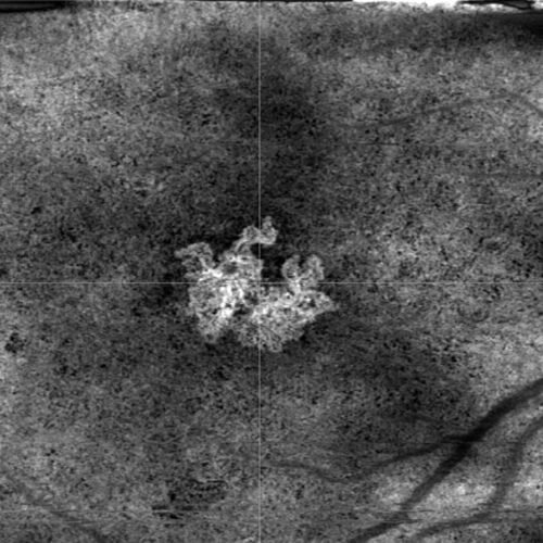 HD 6mm OCT Angiography - Custom sub-RPE segmentation
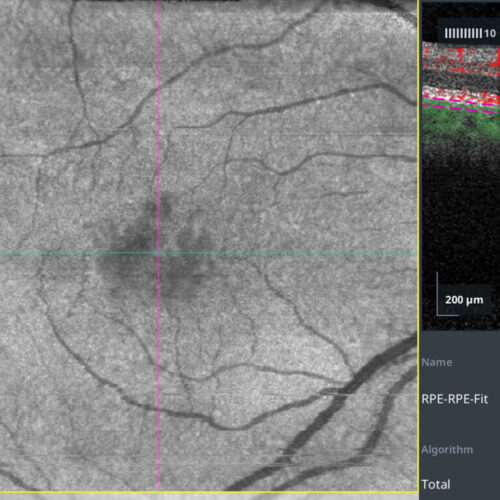 OCT Angiography - Custom sub-RPE segmentation
Quiescent type 1 macular neovascularization (MNV) is a form of neovascular AMD without exudation or hemorrhage. Detection using OCT Angiography (OCTA) involves identifying abnormal blood ves...
Quiescent type 1 macular neovascularization (MNV) is a form of neovascular AMD without exudation or hemorrhage. Detection using OCT Angiography (OCTA) involves identifying abnormal blood ves...
Quiescent type 1 macular neovascularization (MNV) is a form of neovascular AMD without exudation or hemorrhage. Detection using OCT Angiography (OCTA) involves identifying abnormal blood vessels in the sub-RPE space without fluid accumulation. OCTA allows for non-invasive visualization and monitoring, crucial for timely intervention before the onset of exudative changes.
#retina #oftalmo #ophthalmology #oftalmologia #oftalmología #ophtalmologie #офтальмологія #офтальмология #οφθαλμολογία #retinography2024 #CIRRUS6000 #CLARUS700 #ZEISSRETINAWORKFLOW
BackRead More -
Retinitis pigmentosa
Ricardo Leitão Guerra, MD 41yo 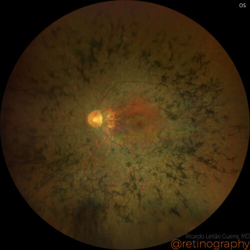 True color
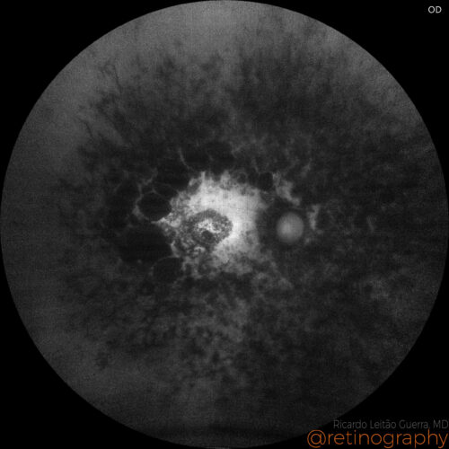 FAF - Green
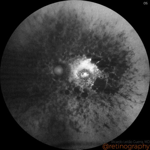 FAF - Green
Retinitis pigmentosa (RP) is a progressive genetic disorder characterized by the degeneration of photoreceptor cells, primarily affecting night and peripheral vision. Fundus autofluorescence...
Retinitis pigmentosa (RP) is a progressive genetic disorder characterized by the degeneration of photoreceptor cells, primarily affecting night and peripheral vision. Fundus autofluorescence...
Retinitis pigmentosa (RP) is a progressive genetic disorder characterized by the degeneration of photoreceptor cells, primarily affecting night and peripheral vision. Fundus autofluorescence (FAF) is a key diagnostic tool in monitoring RP, utilizing the natural fluorescence of lipofuscin in the retinal pigment epithelium (RPE) to detect areas of activity and atrophy. FAF imaging helps identify patterns of progression and areas of retinal degeneration, providing crucial insights for assessing disease severity and potential therapeutic responses.
#retina #oftalmo #ophthalmology #oftalmologia #oftalmología #ophtalmologie #офтальмологія #офтальмология #οφθαλμολογία #retinography2024 #CIRRUS6000 #CLARUS700 #ZEISSRETINAWORKFLOW
BackRead More -
Type 1 MNV
Ricardo Leitão Guerra, MD 74yo
74yo 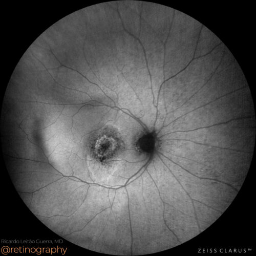 FAF-Green
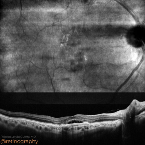 NIR & SD-OCT
Type 1 neovascularization (NVM) in age-related macular degeneration (AMD) involves abnormal blood vessel growth beneath the retinal pigment epithelium. These neovascular membranes are primar...
Type 1 neovascularization (NVM) in age-related macular degeneration (AMD) involves abnormal blood vessel growth beneath the retinal pigment epithelium. These neovascular membranes are primar...
Type 1 neovascularization (NVM) in age-related macular degeneration (AMD) involves abnormal blood vessel growth beneath the retinal pigment epithelium. These neovascular membranes are primarily diagnosed through OCT and fluorescein angiography, showing leakage and progressive vision loss.
#retina #oftalmo #ophthalmology #oftalmologia #oftalmología #ophtalmologie #офтальмологія #офтальмология #οφθαλμολογία #retinography2024 #CIRRUS6000 #CLARUS700 #ZEISSRETINAWORKFLOW
Disclosure: All images featured in this post were acquired and analyzed using devices integrated within the Zeiss Retina Workflow. This ensures high-quality, detailed visual data for comprehensive assessment.
BackRead More -
Valsalva Retinopathy
Ricardo Leitão Guerra, MD 29yo 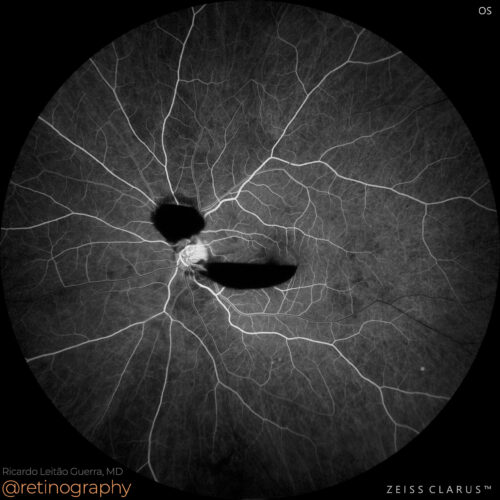 FA: Early phase
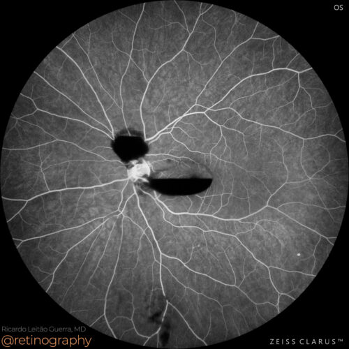 FA: Late phase
 FAF-Green
Valsalva retinopathy is a condition where sudden increases in intraocular venous pressure lead to retinal capillary rupture, causing preretinal hemorrhages. Fluorescein angiography in these ...
Valsalva retinopathy is a condition where sudden increases in intraocular venous pressure lead to retinal capillary rupture, causing preretinal hemorrhages. Fluorescein angiography in these ...
Valsalva retinopathy is a condition where sudden increases in intraocular venous pressure lead to retinal capillary rupture, causing preretinal hemorrhages. Fluorescein angiography in these cases typically shows an absence of fluorescein leakage, which helps differentiate it from other causes of hemorrhage.
#retina #oftalmo #ophthalmology #oftalmologia #oftalmología #ophtalmologie #офтальмологія #офтальмология #οφθαλμολογία #retinography2024 #CIRRUS6000 #CLARUS700 #ZEISSRETINAWORKFLOW
Disclosure: All images featured in this post were acquired and analyzed using devices integrated within the Zeiss Retina Workflow. This ensures high-quality, detailed visual data for comprehensive assessment.
BackRead More -
DONFL
Ricardo Leitão Guerra, MD 78yo 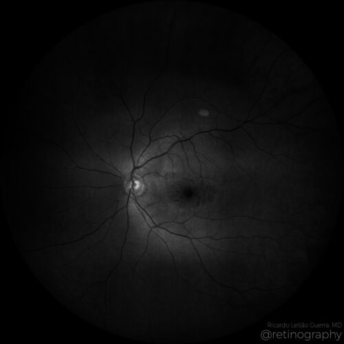 Blue channel
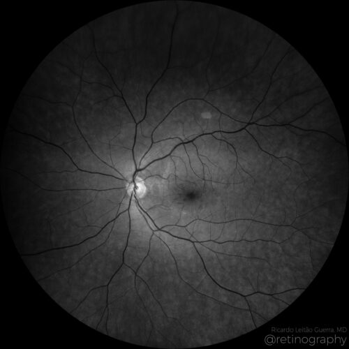 Green channel
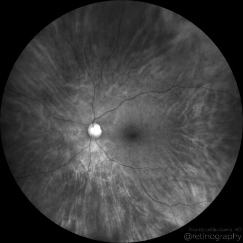 Red channel
Multimodal imaging analysis in a a case of Dissociated Optic Nerve Fiber Layer (DONFL) appearance after internal limiting membrane peeling for idiopathic macular hole. The DONFL appears afte...
Multimodal imaging analysis in a a case of Dissociated Optic Nerve Fiber Layer (DONFL) appearance after internal limiting membrane peeling for idiopathic macular hole. The DONFL appears afte...
Multimodal imaging analysis in a a case of Dissociated Optic Nerve Fiber Layer (DONFL) appearance after internal limiting membrane peeling for idiopathic macular hole. The DONFL appears after vitrectomy, showing as slit-like, arcuate defects in the retinal nerve fiber layer, particularly visible using Optical Coherence Tomography (OCT). The analysis through different color channels enhances visualization, aiding in distinguishing these alterations from other retinal pathologies. Understanding the impact of color channel adjustments is critical for accurate assessment and documentation of DONFL in post-surgical patients.
Disclosure: All images featured in this post were acquired and analyzed using devices integrated within the Zeiss Retina Workflow. This ensures high-quality, detailed visual data for comprehensive assessment.
BackRead More -
Acquired vitelliform lesion
Ricardo Leitão Guerra, MD 92yo 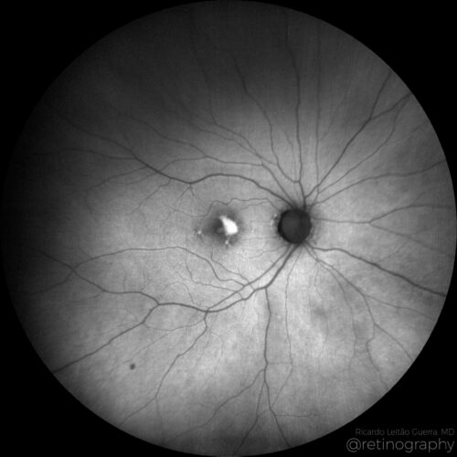 FAF Green
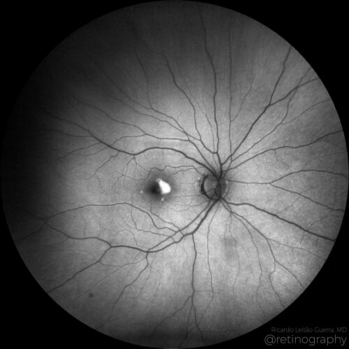 FAF Blue
Acquired vitelliform lesions (AVL) linked to Age-related Macular Degeneration (AMD) present as yellowish round subretinal deposits detectable on fundus examination. Fundus Autofluorescence (...
Acquired vitelliform lesions (AVL) linked to Age-related Macular Degeneration (AMD) present as yellowish round subretinal deposits detectable on fundus examination. Fundus Autofluorescence (...
Acquired vitelliform lesions (AVL) linked to Age-related Macular Degeneration (AMD) present as yellowish round subretinal deposits detectable on fundus examination. Fundus Autofluorescence (FAF) imaging is crucial for AVL detection, showing increased autofluorescence due to lipofuscin accumulation in retinal pigment epithelial cells. These lesions differ from typical AMD deposits and can signify an advanced AMD stage. Understanding AVL’s FAF characteristics helps in differential diagnosis and monitoring AMD progression.
Disclosure: All images featured in this post were acquired and analyzed using devices integrated within the Zeiss Retina Workflow. This ensures high-quality, detailed visual data for comprehensive assessment.
BackRead More -
Acute CSC
Ricardo Leitão Guerra, MD 38yo 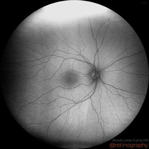 FAF-Blue
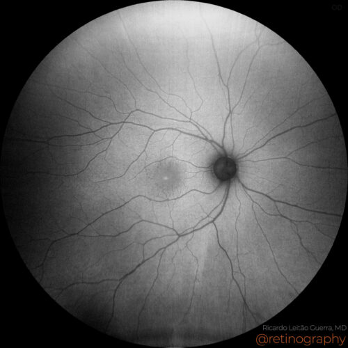 FAF-Green
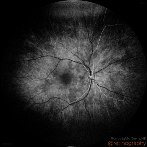 FA: Early phase
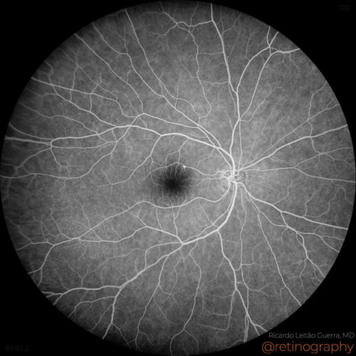 FA: Mid phase
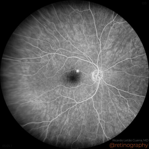 FA: Late phase
Acute central serous chorioretinopathy (CSC) typically presents with serous retinal detachment, primarily affecting the central macula. Key imaging tools include OCT, showing fluid accumulat...
Acute central serous chorioretinopathy (CSC) typically presents with serous retinal detachment, primarily affecting the central macula. Key imaging tools include OCT, showing fluid accumulat...
Acute central serous chorioretinopathy (CSC) typically presents with serous retinal detachment, primarily affecting the central macula. Key imaging tools include OCT, showing fluid accumulation under the retina, and fluorescein angiography, which often reveals a “smokestack” or “inkblot” leakage pattern. Most cases resolve spontaneously, but monitoring is essential.
Disclosure: All images featured in this post were acquired and analyzed using devices integrated within the Zeiss Retina Workflow. This ensures high-quality, detailed visual data for comprehensive assessment.
BackRead More -
Aneurysmal type 1 neovascularization
Ricardo Leitão Guerra, MD 89yo 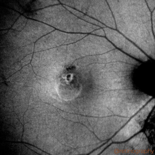 FAF-Green
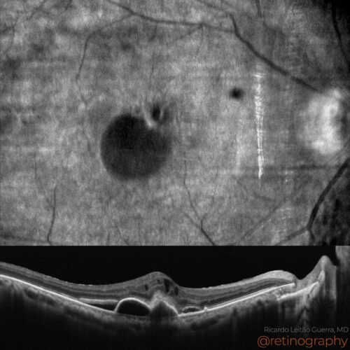 NIR & SD-OCT
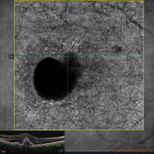 En-face OCT: Structure
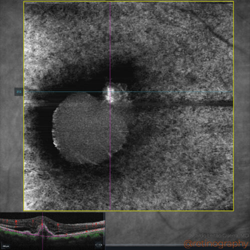 En-face OCT:Angiography
Aneurysmal type 1 neovascularization refers to a subtype of choroidal neovascularization characterized by polypoidal lesions. It is typically identified using OCT and ICGA, showing branching...
Aneurysmal type 1 neovascularization refers to a subtype of choroidal neovascularization characterized by polypoidal lesions. It is typically identified using OCT and ICGA, showing branching...
Aneurysmal type 1 neovascularization refers to a subtype of choroidal neovascularization characterized by polypoidal lesions. It is typically identified using OCT and ICGA, showing branching vascular networks with aneurysmal dilations. This condition may require anti-VEGF therapy and sometimes photodynamic therapy, particularly in treatment-resistant cases.
Disclosure: All images featured in this post were acquired and analyzed using devices integrated within the Zeiss Retina Workflow. This ensures high-quality, detailed visual data for comprehensive assessment.
BackRead More