


Tag:
neovascular AMD
-
AMD: Outer retinal tubulations
Ricardo Leitão Guerra, MD 77yo
77yo  FAF-Green
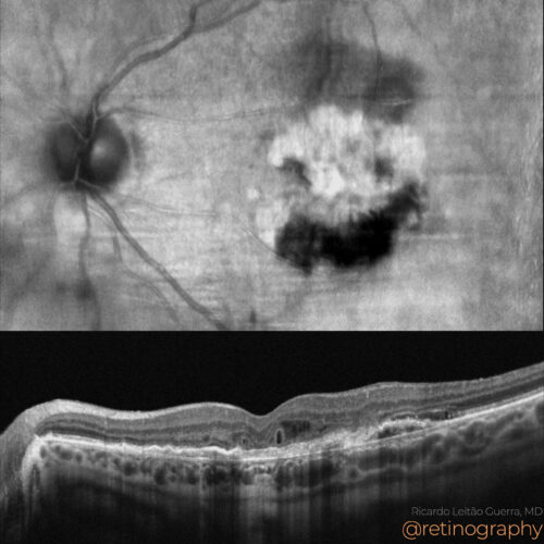 NIR & SD-OCT
NIR & SD-OCT
NIR & SD-OCTIn age-related macular degeneration (AMD), outer retinal tubulations (ORT) can present unique patterns on en-face OCT, often resembling intraretinal fluid (IRF), leading to potential misdiag...
In age-related macular degeneration (AMD), outer retinal tubulations (ORT) can present unique patterns on en-face OCT, often resembling intraretinal fluid (IRF), leading to potential misdiag...
In age-related macular degeneration (AMD), outer retinal tubulations (ORT) can present unique patterns on en-face OCT, often resembling intraretinal fluid (IRF), leading to potential misdiagnosis. In the presented case, en-face imaging shows ORT as hyporreflective interconnected branching networks extensions surrounded by a hyperreflective borders, while dark areas without hyperreflective borders at the periphery represent subretinal fluid (SRF). Accurate interpretation of en-face OCT is essential to differentiate these findings and guide appropriate management.
#AMD #OuterRetinalTubulations #EnFaceOCT #ORT #SubretinalFluid #RetinaImaging #IntraretinalFluid #retina #oftalmo #ophthalmology #oftalmologia #oftalmología #ophtalmologie #офтальмологія #офтальмология #οφθαλμολογία #CIRRUS6000 #CLARUS700 #ZEISSRETINAWORKFLOW
BackRead More -
AMD: Disciform scar
Ricardo Leitão Guerra, MD 77yo 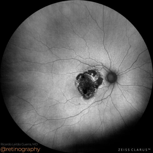 FAF-Green
 NIR & SD-OCT
In advanced age-related macular degeneration (AMD), a disciform scar represents the end stage of neovascular AMD. Optical Coherence Tomography (OCT) shows hyperreflective fibrotic tissue rep...
In advanced age-related macular degeneration (AMD), a disciform scar represents the end stage of neovascular AMD. Optical Coherence Tomography (OCT) shows hyperreflective fibrotic tissue rep...
In advanced age-related macular degeneration (AMD), a disciform scar represents the end stage of neovascular AMD. Optical Coherence Tomography (OCT) shows hyperreflective fibrotic tissue replacing normal retinal layers, with possible subretinal fluid or retinal thinning. Fundus autofluorescence (FAF) reveals hypoautofluorescence due to retinal pigment epithelium (RPE) atrophy, with surrounding hyperautofluorescence indicating RPE stress. These modalities help document structural damage and guide management.
#AMD #DisciformScar #OCT #FAF #RPEAtrophy #RetinaImaging #retina #oftalmo #ophthalmology #oftalmologia #oftalmología #ophtalmologie #офтальмологія #офтальмология #οφθαλμολογία #retinography2024 #CIRRUS6000 #CLARUS700 #ZEISSRETINAWORKFLOW
BackRead More -
AMD: Type 3 MNV
Ricardo Leitão Guerra, MD 74  FAF-Green
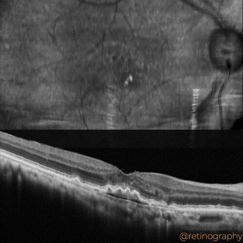 NIR & SD-OCT
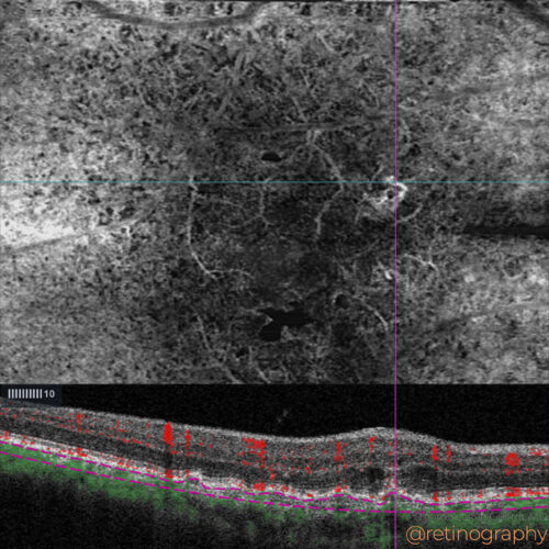 OCT-Angiography
In Type 3 macular neovascularization (MNV), OCT-Angiography B-scan with decorrelation signal is essential for detecting intraretinal neovascularization. The decorrelation signal appears as f...
In Type 3 macular neovascularization (MNV), OCT-Angiography B-scan with decorrelation signal is essential for detecting intraretinal neovascularization. The decorrelation signal appears as f...
In Type 3 macular neovascularization (MNV), OCT-Angiography B-scan with decorrelation signal is essential for detecting intraretinal neovascularization. The decorrelation signal appears as flow signals within the retinal layers on OCT-A, indicating abnormal blood vessels and helping to confirm the presence of Type 3 MNV. This non-invasive imaging technique is critical for diagnosis and monitoring of disease progression.
#Type3MNV #OCTA #BScan #DecorrelacaoSignal #RetinaImaging #retina #oftalmo #ophthalmology #oftalmologia #oftalmología #ophtalmologie #офтальмологія #офтальмология #οφθαλμολογία #retinography2024 #CIRRUS6000 #CLARUS700 #ZEISSRETINAWORKFLOW
BackRead More -
AMD: RPE tear
Ricardo Leitão Guerra, MD 78yo 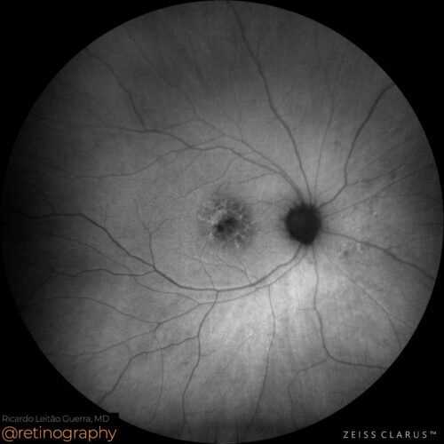 FAF-Green
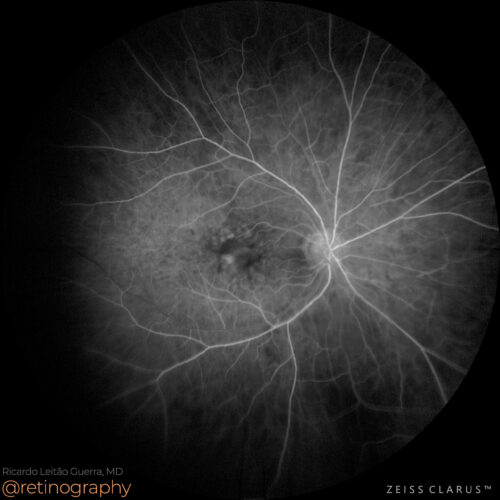 FA: Early phase
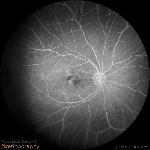 FA: Mid phase
 FA: Late phase
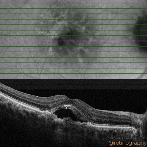 FAF-Green & SD-OCT
In age-related macular degeneration (AMD), a retinal pigment epithelium (RPE) tear is a serious complication often associated with pigment epithelial detachments. Fundus autofluorescence (FA...
In age-related macular degeneration (AMD), a retinal pigment epithelium (RPE) tear is a serious complication often associated with pigment epithelial detachments. Fundus autofluorescence (FA...
In age-related macular degeneration (AMD), a retinal pigment epithelium (RPE) tear is a serious complication often associated with pigment epithelial detachments. Fundus autofluorescence (FAF) shows hypoautofluorescence in the area of the tear due to the loss of RPE, while hyperautofluorescence may outline the borders. Optical Coherence Tomography (OCT) provides cross-sectional images, revealing the extent of the RPE tear and associated subretinal fluid, aiding in diagnosis and monitoring.
#AMD #RPETear #FAF #OCT #RetinaImaging #retina #oftalmo #ophthalmology #oftalmologia #oftalmología #ophtalmologie #офтальмологія #офтальмология #οφθαλμολογία #retinography2024 #CIRRUS6000 #CLARUS700 #ZEISSRETINAWORKFLOW
BackRead More -
Neovascular AMD
Ricardo Leitão Guerra, MD 90 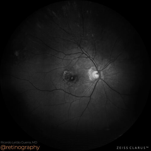 Blue channel
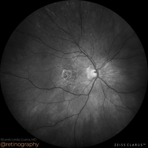 Green channel
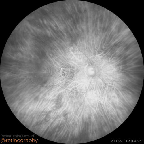 Red channel
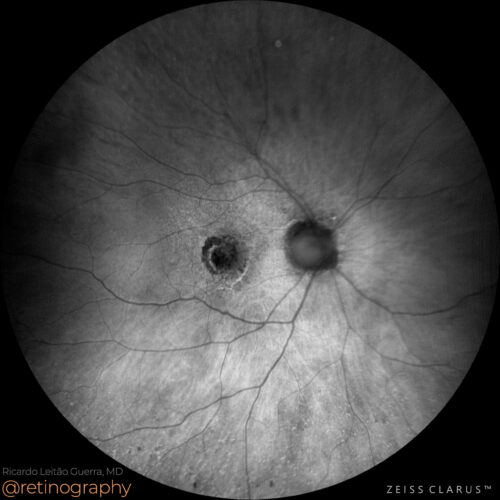 FAF-Green
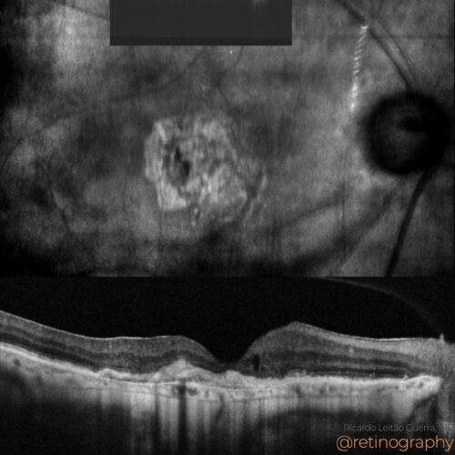 NIR & SD-OCT
In neovascular age-related macular degeneration (AMD), multimodal evaluation is essential for accurate diagnosis and management. Optical Coherence Tomography (OCT) detects subretinal and int...
In neovascular age-related macular degeneration (AMD), multimodal evaluation is essential for accurate diagnosis and management. Optical Coherence Tomography (OCT) detects subretinal and int...
In neovascular age-related macular degeneration (AMD), multimodal evaluation is essential for accurate diagnosis and management. Optical Coherence Tomography (OCT) detects subretinal and intraretinal fluid, indicating neovascular activity. Fundus autofluorescence (FAF) helps assess retinal pigment epithelium (RPE) status. Combining these modalities provides a comprehensive view of the disease for optimal treatment planning.
#NeovascularAMD #MultimodalEvaluation #OCT #OCTA #FAF #FluoresceinAngiography #RetinaImaging #retina #oftalmo #ophthalmology #oftalmologia #oftalmología #ophtalmologie #офтальмологія #офтальмология #οφθαλμολογία #retinography2024 #CIRRUS6000 #CLARUS700 #ZEISSRETINAWORKFLOW
BackRead More -
Neovascular AMD
Ricardo Leitão Guerra, MD 71yo 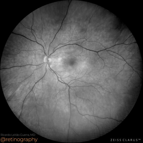 Green channel
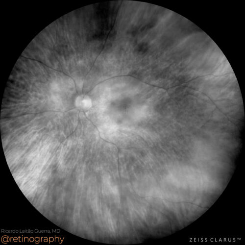 Red channel
 FAF-Green
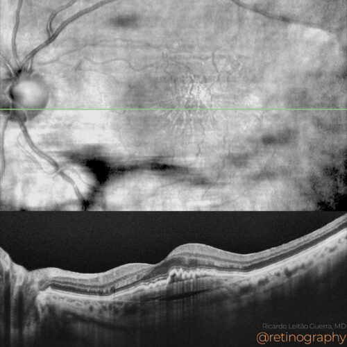 NIR & SD-OCT
 OCT-Angiography
In neovascular age-related macular degeneration (AMD) with Type 1 macular neovascularization (MNV), a prechoroidal cleft may be observed. This cleft appears as a hyporeflective space between...
In neovascular age-related macular degeneration (AMD) with Type 1 macular neovascularization (MNV), a prechoroidal cleft may be observed. This cleft appears as a hyporeflective space between...
In neovascular age-related macular degeneration (AMD) with Type 1 macular neovascularization (MNV), a prechoroidal cleft may be observed. This cleft appears as a hyporeflective space between the neovascular membrane and the choroid on Optical Coherence Tomography (OCT). Identifying this feature is crucial for accurate diagnosis and management, guiding treatment with anti-VEGF therapy to address the neovascular activity.
#NeovascularAMD #Type1MNV #PrechoroidalCleft #OCT #AntiVEGF #RetinaImaging #retina #oftalmo #ophthalmology #oftalmologia #oftalmología #ophtalmologie #офтальмологія #офтальмология #οφθαλμολογία #retinography2024 #CIRRUS6000 #CLARUS700 #ZEISSRETINAWORKFLOW
BackRead More -
CME and Neovascular AMD
Ricardo Leitão Guerra, MD 71yo 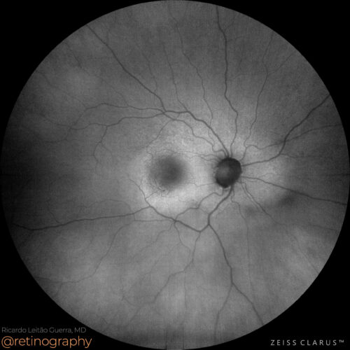 FAF-Green
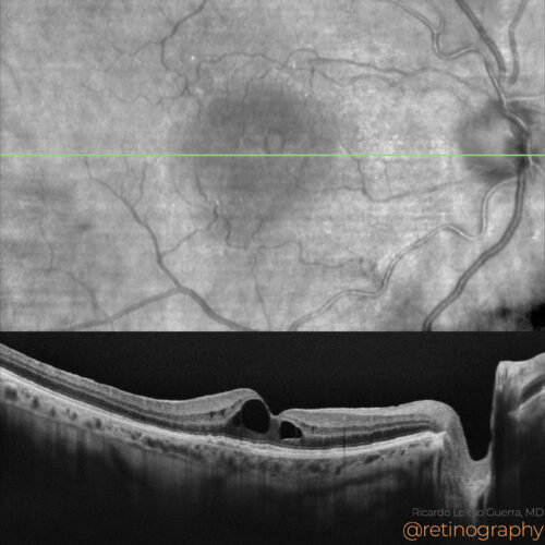 NIR & SD-OCT
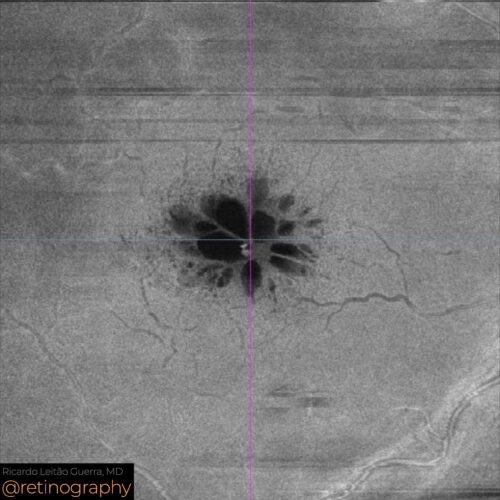 En-face: Mid Retina
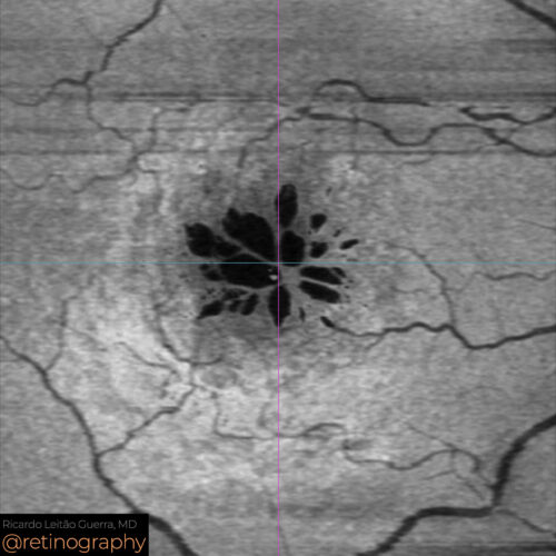 En-face: Minimum intensity
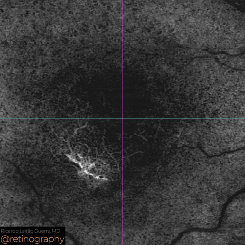 OCT-Angiography
Cystoid macular edema (CME) following phacoemulsification in a patient with neovascular age-related macular degeneration (AMD) and Type 1 neovascular membrane requires careful management. Op...
Cystoid macular edema (CME) following phacoemulsification in a patient with neovascular age-related macular degeneration (AMD) and Type 1 neovascular membrane requires careful management. Op...
Cystoid macular edema (CME) following phacoemulsification in a patient with neovascular age-related macular degeneration (AMD) and Type 1 neovascular membrane requires careful management. Optical Coherence Tomography (OCT) can be used to monitor the edema and the neovascular membrane. Treatment may include anti-VEGF injections and corticosteroids to reduce inflammation and manage the edema.
#CME #NeovascularAMD #Type1NeovascularMembrane #Phacoemulsification #OCT #AntiVEGF #RetinaImaging #retina #oftalmo #ophthalmology #oftalmologia #oftalmología #ophtalmologie #офтальмологія #офтальмология #οφθαλμολογία #retinography2024 #CIRRUS6000 #CLARUS700 #ZEISSRETINAWORKFLOW
BackRead More -
Neovascular AMD
Ricardo Leitão Guerra, MD 77yo 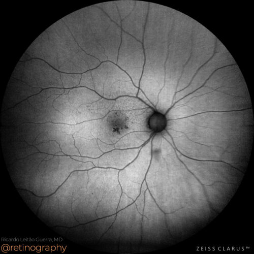 FAF-Green
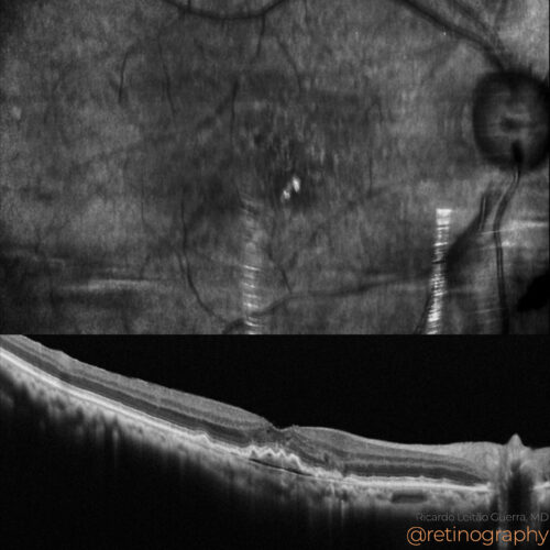 NIR & SD-OCT
 OCT-Angiography
The early stages of neovascular age-related macular degeneration (AMD) can be challenging to diagnose. Optical Coherence Tomography (OCT) and Optical Coherence Tomography Angiography (OCTA) ...
The early stages of neovascular age-related macular degeneration (AMD) can be challenging to diagnose. Optical Coherence Tomography (OCT) and Optical Coherence Tomography Angiography (OCTA) ...
BackRead More -
Neovascular AMD
Ricardo Leitão Guerra, MD 94yo 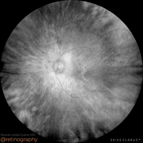 Red channel
 Green channel
 Blue channel
 FAF-Green
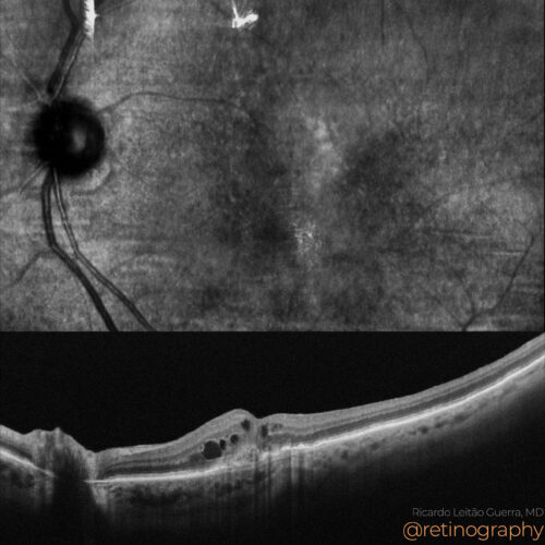 NIR & SD-OCT
 OCT-Angiography
 OCT-Angiography
Neovascular age-related macular degeneration (AMD) often presents with pseudodrusen, which can be highlighted using the blue channel in color fundus photography. Optical Coherence Tomography...
Neovascular age-related macular degeneration (AMD) often presents with pseudodrusen, which can be highlighted using the blue channel in color fundus photography. Optical Coherence Tomography...
BackRead More -
Not just drusen – Type 3 MNV
Ricardo Leitão Guerra, MD 74yo
74yo  FAF-Green
 NIR & SD-OCT
 Screenshot
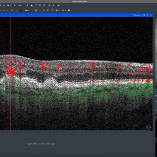 OCT-Angiography
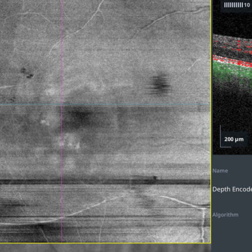 OCT-Angiography
Check out this case: A 73-year-old male presented with apparent non-neovascular age-related macular degeneration (AMD) in the right eye. True color imaging revealed multiple confluent soft d...
Check out this case: A 73-year-old male presented with apparent non-neovascular age-related macular degeneration (AMD) in the right eye. True color imaging revealed multiple confluent soft d...
Check out this case: A 73-year-old male presented with apparent non-neovascular age-related macular degeneration (AMD) in the right eye. True color imaging revealed multiple confluent soft drusen across the macular area. FAF-green imaging showed hypoautofluorescent spots over some of these drusen. SD-OCT B-scan images clearly depicted the drusen as homogeneous, medium-to-high reflectivity elevations of the retinal pigment epithelium (RPE). Advanced RPE analysis enabled precise evaluation of the area and volume of these drusen, facilitating intuitive comparisons over time. However, a more detailed analysis using OCTA revealed type 3 macular neovascularization with adjacent subtle intraretinal fluid. This case underscores the importance of thorough evaluation in AMD patients, utilizing all tools in multimodal retinal imaging for the early detection of more severe forms.
#retina #oftalmo #ophthalmology #oftalmologia #oftalmología #ophtalmologie #офтальмологія #офтальмология #οφθαλμολογία #retinography2024 #CIRRUS6000 #CLARUS700 #ZEISSRETINAWORKFLOW
BackRead More