


Tag:
Rhegmatogenous retinal detachment
-
Rhegmatogenous retinal detachment
Ricardo Leitão Guerra, MD 56yo
56yo 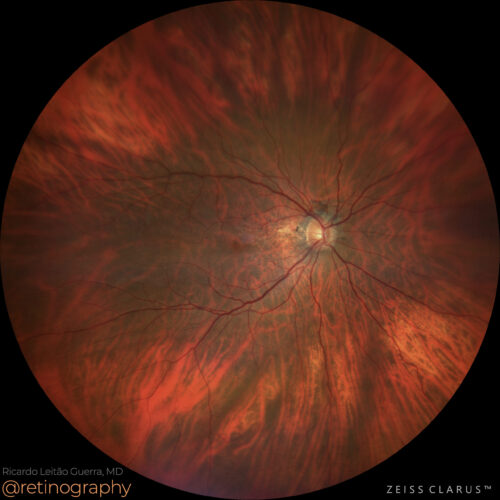 True color - Follow up
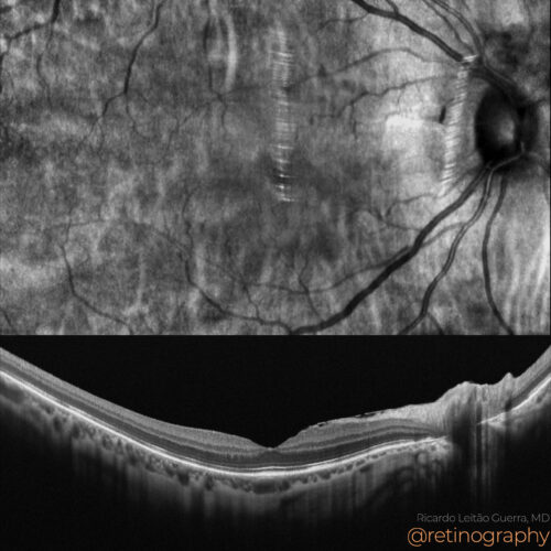 NIR & SD-OCT
In macula-off rhegmatogenous retinal detachment, post-surgical recovery can be evaluated using Optical Coherence Tomography (OCT). An excellent recovery of the external retina is characteriz...
In macula-off rhegmatogenous retinal detachment, post-surgical recovery can be evaluated using Optical Coherence Tomography (OCT). An excellent recovery of the external retina is characteriz...
In macula-off rhegmatogenous retinal detachment, post-surgical recovery can be evaluated using Optical Coherence Tomography (OCT). An excellent recovery of the external retina is characterized by the restoration of the ellipsoid zone (EZ) and external limiting membrane (ELM). These findings on OCT are associated with better visual outcomes, indicating effective surgical intervention and retinal reattachment.
#RhegmatogenousRetinalDetachment #MaculaOff #OCT #ExternalRetinaRecovery #RetinaImaging #retina #oftalmo #ophthalmology #oftalmologia #oftalmología #ophtalmologie #офтальмологія #офтальмология #οφθαλμολογία #retinography2024 #CIRRUS6000 #CLARUS700 #ZEISSRETINAWORKFLOW
BackRead More -
Low-integrity retinal attachment
Ricardo Leitão Guerra, MD 65 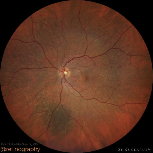 True color
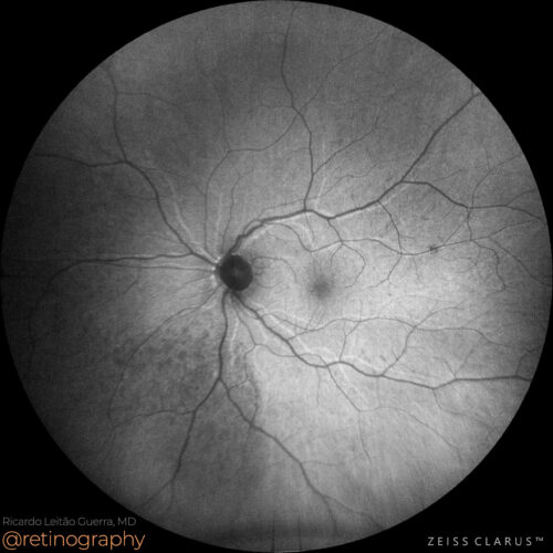 FAF-Green
Retinal reattachment in the wrong position can occur following surgery for retinal detachment, and fundus autofluorescence (FAF) can reveal hyperautofluorescent lines where the retinal vesse...
Retinal reattachment in the wrong position can occur following surgery for retinal detachment, and fundus autofluorescence (FAF) can reveal hyperautofluorescent lines where the retinal vesse...
Retinal reattachment in the wrong position can occur following surgery for retinal detachment, and fundus autofluorescence (FAF) can reveal hyperautofluorescent lines where the retinal vessels originally lay. These lines represent areas of misalignment and retinal pigment epithelium (RPE) stress, providing insight into the surgical outcome and potential functional impact.
#RetinalReattachment #FAF #Hyperautofluorescence #RetinaSurgery #RetinaImaging #retina #oftalmo #ophthalmology #oftalmologia #oftalmología #ophtalmologie #офтальмологія #офтальмология #οφθαλμολογία #retinography2024 #CIRRUS6000 #CLARUS700 #ZEISSRETINAWORKFLOW
BackRead More -
Giant retinal tear detachment
Ricardo Leitão Guerra, MD 46 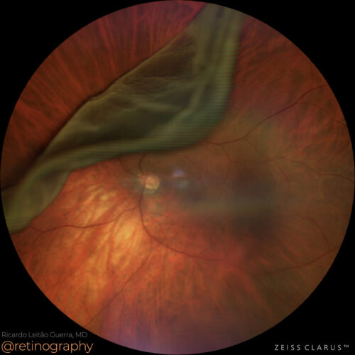 True color
A giant retinal tear is a full-thickness break in the retina, often exceeding 90 degrees of circumference, and frequently leads to retinal detachment. This condition requires urgent interven...
A giant retinal tear is a full-thickness break in the retina, often exceeding 90 degrees of circumference, and frequently leads to retinal detachment. This condition requires urgent interven...
A giant retinal tear is a full-thickness break in the retina, often exceeding 90 degrees of circumference, and frequently leads to retinal detachment. This condition requires urgent intervention, typically with vitrectomy and retinal reattachment surgery, to prevent further vision loss. Early detection and prompt surgical management are crucial for preserving vision.
#GiantRetinalTear #RetinalDetachment #Vitrectomy #RetinaSurgery #RetinaImaging #retina #oftalmo #ophthalmology #oftalmologia #oftalmología #ophtalmologie #офтальмологія #офтальмология #οφθαλμολογία #retinography2024 #CIRRUS6000 #CLARUS700 #ZEISSRETINAWORKFLOW
BackRead More -
RRD: Giant retinal tear
Ricardo Leitão Guerra, MD 51yo 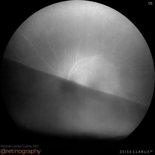 FAF-Green
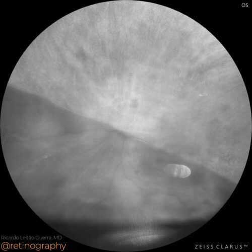 NIR & SD-OCT
Giant retinal tears can be assessed using fundus autofluorescence (FAF), which reveals the previous location of retinal vessels as hyperautofluorescent lines. These lines indicate where the ...
Giant retinal tears can be assessed using fundus autofluorescence (FAF), which reveals the previous location of retinal vessels as hyperautofluorescent lines. These lines indicate where the ...
Giant retinal tears can be assessed using fundus autofluorescence (FAF), which reveals the previous location of retinal vessels as hyperautofluorescent lines. These lines indicate where the retinal vessels were detached from the underlying retinal pigment epithelium, aiding in the evaluation of the extent of the tear and guiding surgical repair.
#GiantRetinalTear #FAF #Hyperautofluorescence #RetinaImaging #retina #oftalmo #ophthalmology #oftalmologia #oftalmología #ophtalmologie #офтальмологія #офтальмология #οφθαλμολογία #retinography2024 #CIRRUS6000 #CLARUS700 #ZEISSRETINAWORKFLOW
BackRead More -
Peripheral degeneration
Ricardo Leitão Guerra, MD 42yo 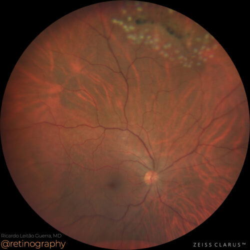 True color
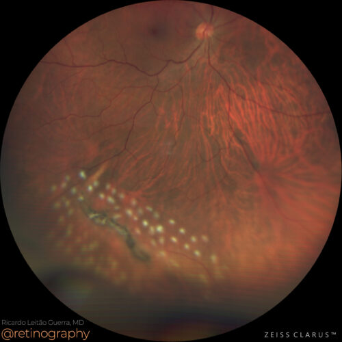 True color
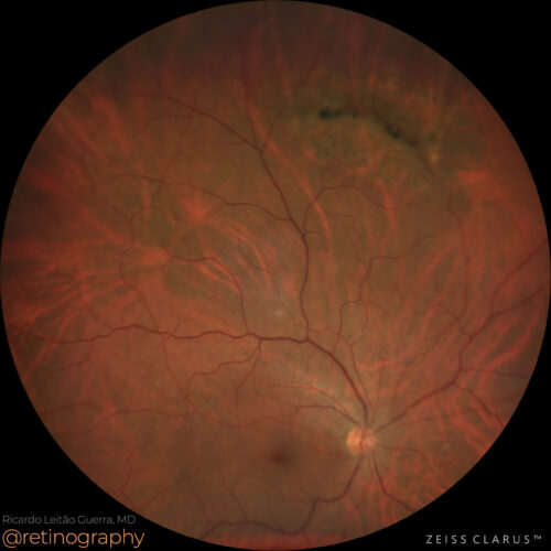 True color: Follow up
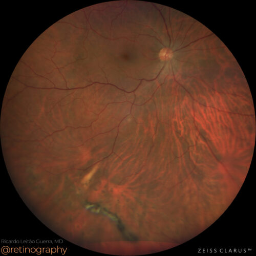 True color: Follow up
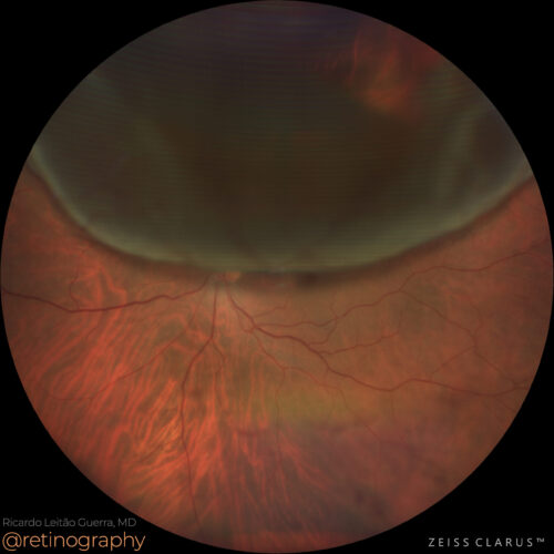 True color: Fellow eye
A 42-year-old man presents with lattice degenerations in the peripheral retina of the right eye. Ultra-widefield imaging perfectly documents these lesions. Due to a history of retinal detach...
A 42-year-old man presents with lattice degenerations in the peripheral retina of the right eye. Ultra-widefield imaging perfectly documents these lesions. Due to a history of retinal detach...
BackRead More -
Degenerative retinoschisis & retinal detachment
Ricardo Leitão Guerra, MD 40yo 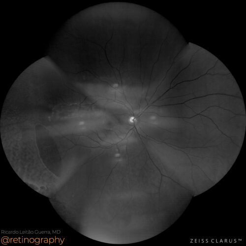 Blue-channel
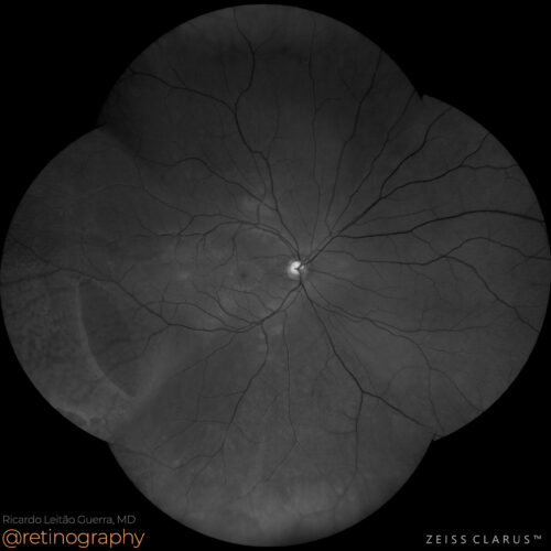 Green-channel
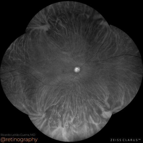 Red-channel
 True color
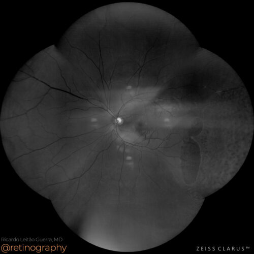 Blue-channel
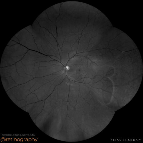 Green-channel
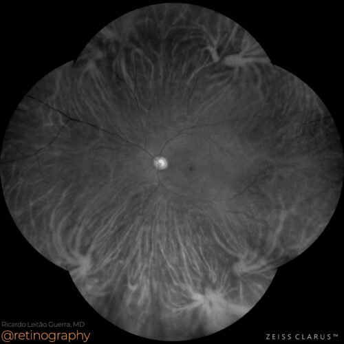 Red-channel
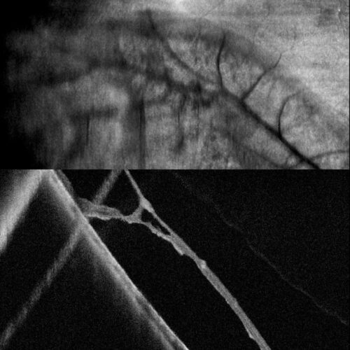 NIR & SD-OCT
Degenerative retinoschisis involves the splitting of retinal layers, which can lead to retinal detachment if a tear occurs in both layers, such as in this case. #retina #oftalmo #ophthalmolo...
Degenerative retinoschisis involves the splitting of retinal layers, which can lead to retinal detachment if a tear occurs in both layers, such as in this case. #retina #oftalmo #ophthalmolo...
Degenerative retinoschisis involves the splitting of retinal layers, which can lead to retinal detachment if a tear occurs in both layers, such as in this case.
#retina #oftalmo #ophthalmology #oftalmologia #oftalmología #ophtalmologie #офтальмологія #офтальмология #οφθαλμολογία #retinography2024 #CIRRUS6000 #CLARUS700 #ZEISSRETINAWORKFLOW
BackRead More -
Rhegmatogenous Retinal Detachment
Ricardo Leitão Guerra, MD 41yo 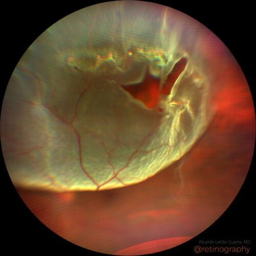 Detached retina - Superior tear
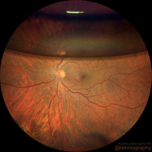 Retina reattached after PPV + C3F8
A 41yo male presented with rhegmatogenous retinal detachment (macula on) and multiple retinal tears, the larger one was superior. He was treated by primary pars plana vitrectomy. Forty days ...
A 41yo male presented with rhegmatogenous retinal detachment (macula on) and multiple retinal tears, the larger one was superior. He was treated by primary pars plana vitrectomy. Forty days ...
A 41yo male presented with rhegmatogenous retinal detachment (macula on) and multiple retinal tears, the larger one was superior. He was treated by primary pars plana vitrectomy. Forty days after surgery BCVA was 20/20 and C3F8 was filling 1/3 of the vitreous cavity. Vitrectomy with gas injection is a common treatment for retinal detachment, involving the removal of the vitreous gel and injecting a gas bubble to press the retina back into place. The gas bubble helps flatten the retina against the wall of the eye, facilitating reattachment and healing.
Disclosure: All images featured in this post were acquired and analyzed using devices integrated within the Zeiss Retina Workflow. This ensures high-quality, detailed visual data for comprehensive assessment.
BackRead More