


Tag:
Type 1 macular neovascularization
-
Neovascular AMD
Ricardo Leitão Guerra, MD 71yo
71yo 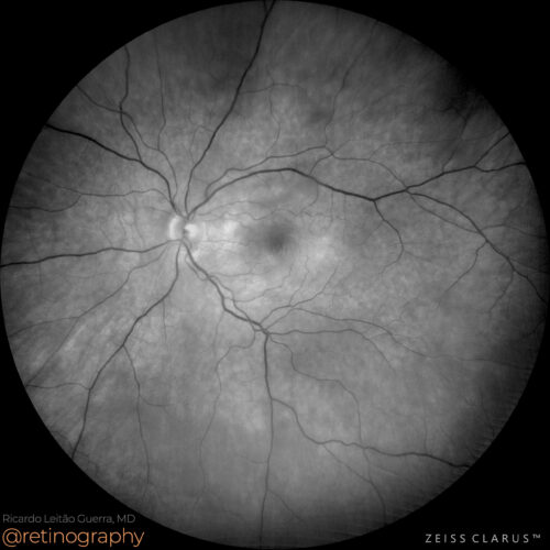 Green channel
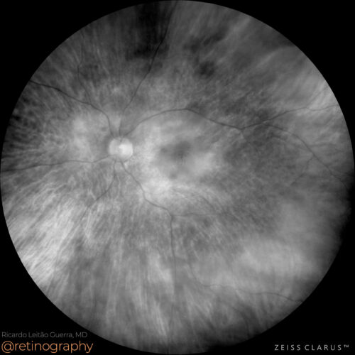 Red channel
 FAF-Green
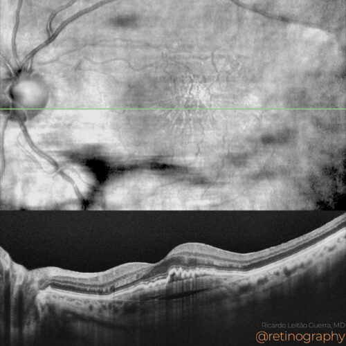 NIR & SD-OCT
 OCT-Angiography
In neovascular age-related macular degeneration (AMD) with Type 1 macular neovascularization (MNV), a prechoroidal cleft may be observed. This cleft appears as a hyporeflective space between...
In neovascular age-related macular degeneration (AMD) with Type 1 macular neovascularization (MNV), a prechoroidal cleft may be observed. This cleft appears as a hyporeflective space between...
In neovascular age-related macular degeneration (AMD) with Type 1 macular neovascularization (MNV), a prechoroidal cleft may be observed. This cleft appears as a hyporeflective space between the neovascular membrane and the choroid on Optical Coherence Tomography (OCT). Identifying this feature is crucial for accurate diagnosis and management, guiding treatment with anti-VEGF therapy to address the neovascular activity.
#NeovascularAMD #Type1MNV #PrechoroidalCleft #OCT #AntiVEGF #RetinaImaging #retina #oftalmo #ophthalmology #oftalmologia #oftalmología #ophtalmologie #офтальмологія #офтальмология #οφθαλμολογία #retinography2024 #CIRRUS6000 #CLARUS700 #ZEISSRETINAWORKFLOW
BackRead More -
CME and Neovascular AMD
Ricardo Leitão Guerra, MD 71yo 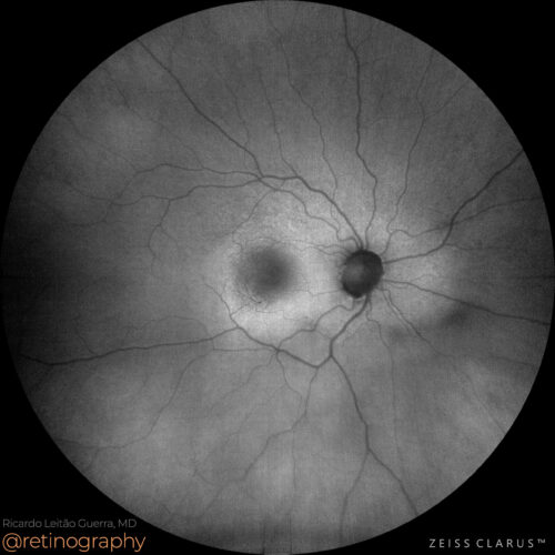 FAF-Green
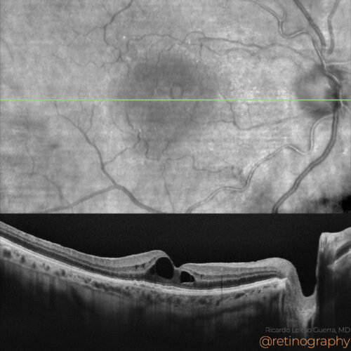 NIR & SD-OCT
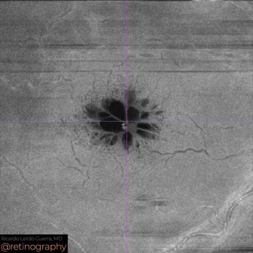 En-face: Mid Retina
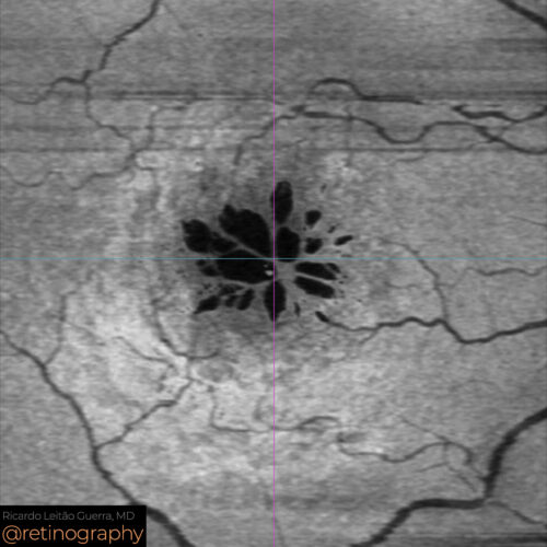 En-face: Minimum intensity
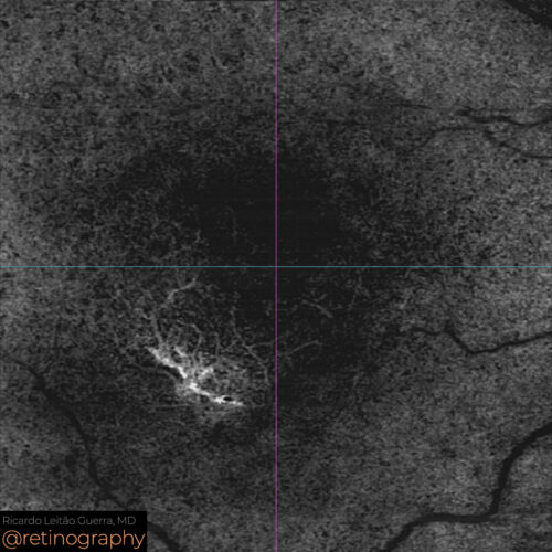 OCT-Angiography
Cystoid macular edema (CME) following phacoemulsification in a patient with neovascular age-related macular degeneration (AMD) and Type 1 neovascular membrane requires careful management. Op...
Cystoid macular edema (CME) following phacoemulsification in a patient with neovascular age-related macular degeneration (AMD) and Type 1 neovascular membrane requires careful management. Op...
Cystoid macular edema (CME) following phacoemulsification in a patient with neovascular age-related macular degeneration (AMD) and Type 1 neovascular membrane requires careful management. Optical Coherence Tomography (OCT) can be used to monitor the edema and the neovascular membrane. Treatment may include anti-VEGF injections and corticosteroids to reduce inflammation and manage the edema.
#CME #NeovascularAMD #Type1NeovascularMembrane #Phacoemulsification #OCT #AntiVEGF #RetinaImaging #retina #oftalmo #ophthalmology #oftalmologia #oftalmología #ophtalmologie #офтальмологія #офтальмология #οφθαλμολογία #retinography2024 #CIRRUS6000 #CLARUS700 #ZEISSRETINAWORKFLOW
BackRead More -
PCV: Quiescent MNV
Ricardo Leitão Guerra, MD 64yo 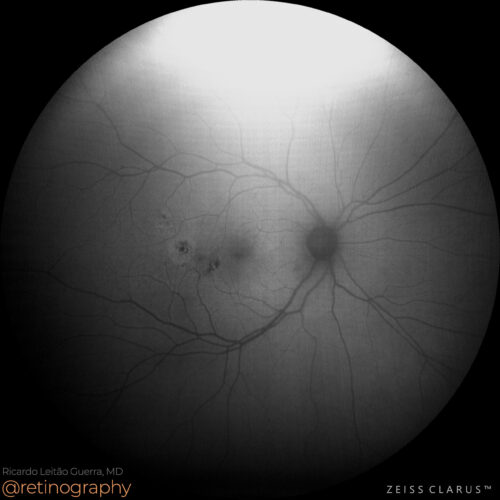 FAF-Green
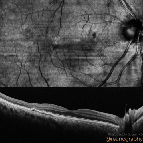 NIR & SD-OCT
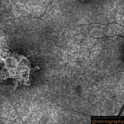 OCT-Angiography
Quiescent Type 1 macular neovascularization (MNV) and polypoidal choroidal vasculopathy (PCV) can be assessed using Optical Coherence Tomography Angiography (OCTA). OCTA provides detailed im...
Quiescent Type 1 macular neovascularization (MNV) and polypoidal choroidal vasculopathy (PCV) can be assessed using Optical Coherence Tomography Angiography (OCTA). OCTA provides detailed im...
Quiescent Type 1 macular neovascularization (MNV) and polypoidal choroidal vasculopathy (PCV) can be assessed using Optical Coherence Tomography Angiography (OCTA). OCTA provides detailed images of the vascular structures, allowing for the identification and monitoring of these conditions without the need for dye injection, thereby facilitating non-invasive diagnosis and management.
#Type1MNV #PCV #OCTA #RetinaImaging #retina #oftalmo #ophthalmology #oftalmologia #oftalmología #ophtalmologie #офтальмологія #офтальмология #οφθαλμολογία #retinography2024 #CIRRUS6000 #CLARUS700 #ZEISSRETINAWORKFLOW
BackRead More -
Choroidal nevus
Ricardo Leitão Guerra, MD 51yo
51yo 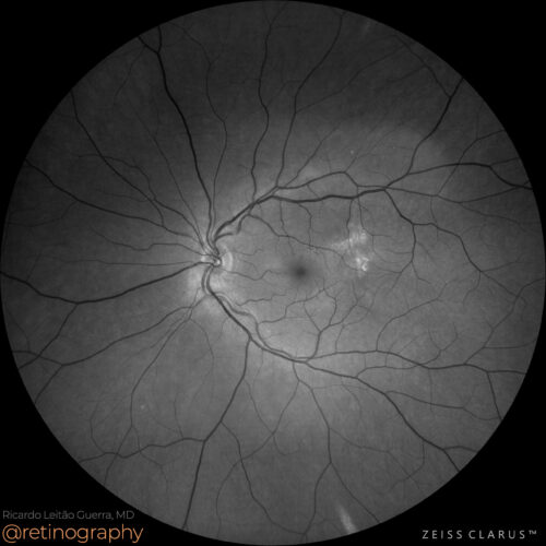 Green channel
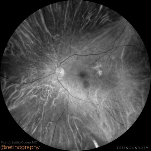 Red channel
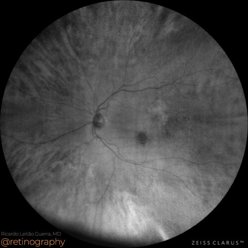 NIR
NIR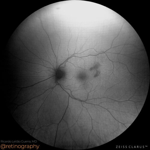 FAF-Green
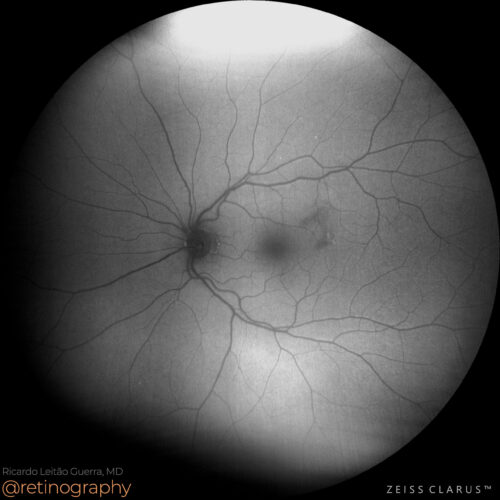 FAF-Blue
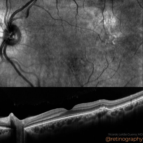 MODELO SITE DIESSY.008
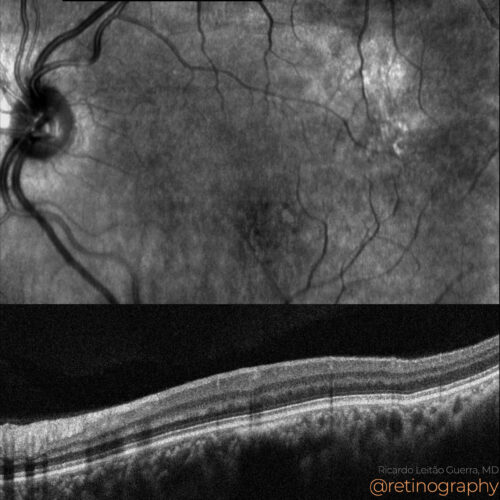 NIR & SD-OCT
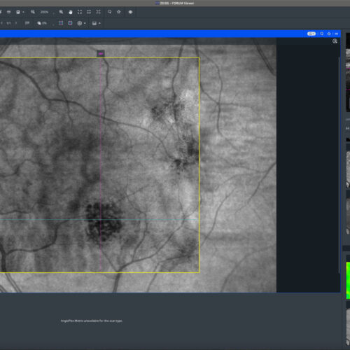 En-face: Choroid
En-face: Choroid
En-face: ChoroidChoroidal nevus is a benign pigmented lesion in the choroid, often monitored for potential malignant transformation. Near-Infrared Reflectance (NIR) imaging and the red channel in color fund...
Choroidal nevus is a benign pigmented lesion in the choroid, often monitored for potential malignant transformation. Near-Infrared Reflectance (NIR) imaging and the red channel in color fund...
Choroidal nevus is a benign pigmented lesion in the choroid, often monitored for potential malignant transformation. Near-Infrared Reflectance (NIR) imaging and the red channel in color fundus photography enhance the visibility of the nevus. Optical Coherence Tomography (OCT) provides detailed cross-sectional images to assess the nevus’s impact on retinal layers. OCT Angiography (OCTA) evaluates the blood flow around the lesion, aiding in detecting any neovascular activity. In this case there is also a quiescent type 1 macular neovascularization at the temporal-superior macular area.
#retina #oftalmo #ophthalmology #oftalmologia #oftalmología #ophtalmologie #офтальмологія #офтальмология #οφθαλμολογία #retinography2024 #CIRRUS6000 #CLARUS700 #ZEISSRETINAWORKFLOW
BackRead More -
MNV Type-1 and RPE Tear
Ricardo Leitão Guerra, MD 79yo 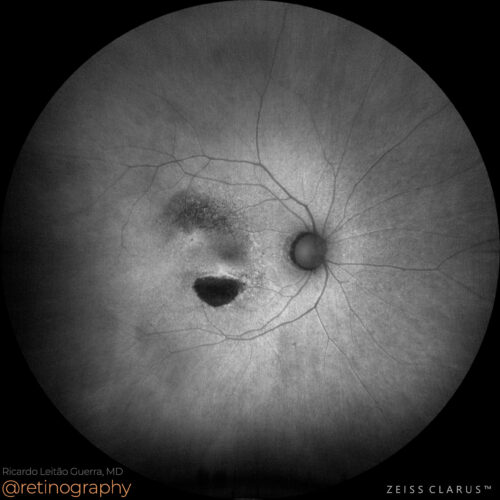 FAF-Green
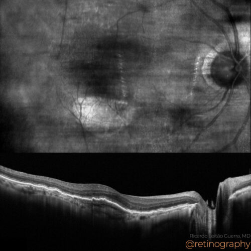 NIR & SD-OCT
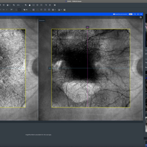 OCT-Angiography
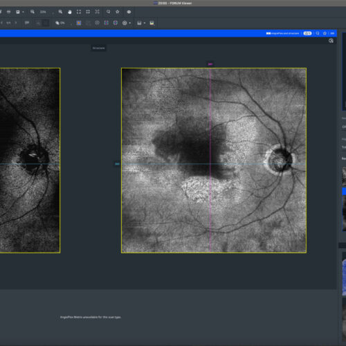 OCT-Angiography
 En-face: Choroid
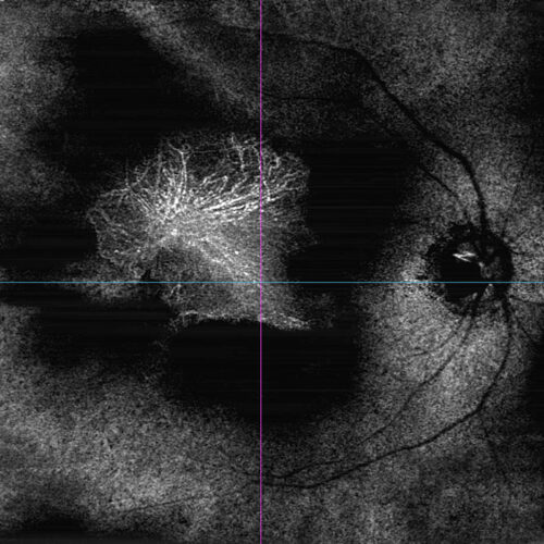 OCT-Angiography
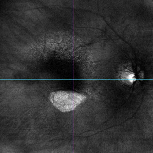 En-face: Choroid
Type 1 macular neovascularization (MNV), commonly associated with subretinal pigment epithelium (RPE) growth, may not be fully visualized on standard 6×6 mm OCTA scans. However, wide-fi...
Type 1 macular neovascularization (MNV), commonly associated with subretinal pigment epithelium (RPE) growth, may not be fully visualized on standard 6×6 mm OCTA scans. However, wide-fi...
Type 1 macular neovascularization (MNV), commonly associated with subretinal pigment epithelium (RPE) growth, may not be fully visualized on standard 6×6 mm OCTA scans. However, wide-field OCTA, with its 12×12 mm view, captures the entire extent of these lesions, providing a comprehensive assessment of the neovascular complex and its spread beyond the central macula. Fundus autofluorescence (FAF) and en-face OCT hyper-transmission imaging are effective tools for assessing retinal pigment epithelium (RPE) tears. FAF highlights areas of RPE loss or damage by showing increased autofluorescence, while en-face OCT reveals hyper-transmission below the tear due to enhanced light penetration. Together, they provide detailed insights into the extent and impact of RPE disruption.
#retina #oftalmo #ophthalmology #oftalmologia #oftalmología #ophtalmologie #офтальмологія #офтальмология #οφθαλμολογία #retinography2024 #CIRRUS6000 #CLARUS700 #ZEISSRETINAWORKFLOW
BackRead More -
AMD – Type 1 MNV
Ricardo Leitão Guerra, MD 83yo 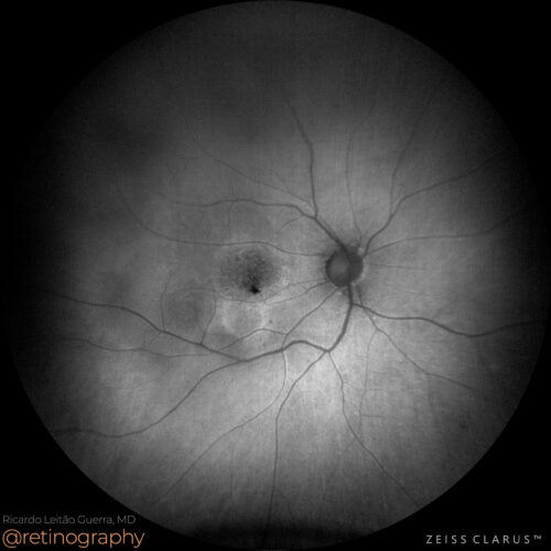 FAF-Green
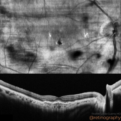 NIR & SD-OCT
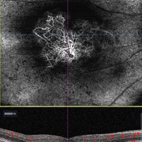 OCT-Angiography: RPE - RPE fit
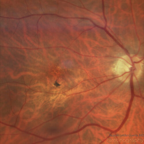 True color - Zoom
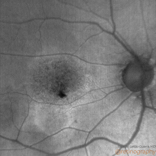 MODELO SITE DIESSY.025
Type 1 macular neovascularization (MNV) involves the growth of abnormal blood vessels beneath the retinal pigment epithelium, often associated with age-related macular degeneration. Optical ...
Type 1 macular neovascularization (MNV) involves the growth of abnormal blood vessels beneath the retinal pigment epithelium, often associated with age-related macular degeneration. Optical ...
Type 1 macular neovascularization (MNV) involves the growth of abnormal blood vessels beneath the retinal pigment epithelium, often associated with age-related macular degeneration. Optical coherence tomography angiography (OCT-A) is crucial for detecting and monitoring Type 1 MNV, providing detailed images of blood flow and vascular structure without the need for dye injection. This non-invasive technique aids in early diagnosis and management.
#retina #oftalmo #ophthalmology #oftalmologia #oftalmología #ophtalmologie #офтальмологія #офтальмология #οφθαλμολογία #retinography2024 #CIRRUS6000 #CLARUS700 #ZEISSRETINAWORKFLOW
BackRead More -
Quiescent type 1 MNV
Ricardo Leitão Guerra, MD 80yo 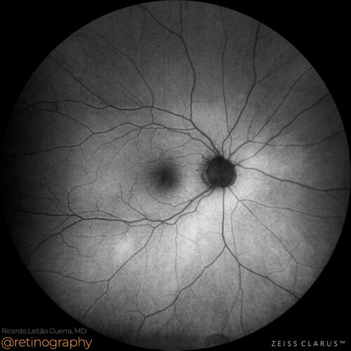 FAF-Green
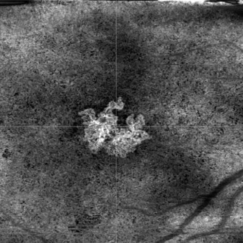 HD 6mm OCT Angiography - Custom sub-RPE segmentation
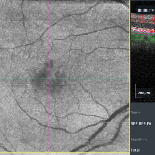 OCT Angiography - Custom sub-RPE segmentation
Quiescent type 1 macular neovascularization (MNV) is a form of neovascular AMD without exudation or hemorrhage. Detection using OCT Angiography (OCTA) involves identifying abnormal blood ves...
Quiescent type 1 macular neovascularization (MNV) is a form of neovascular AMD without exudation or hemorrhage. Detection using OCT Angiography (OCTA) involves identifying abnormal blood ves...
Quiescent type 1 macular neovascularization (MNV) is a form of neovascular AMD without exudation or hemorrhage. Detection using OCT Angiography (OCTA) involves identifying abnormal blood vessels in the sub-RPE space without fluid accumulation. OCTA allows for non-invasive visualization and monitoring, crucial for timely intervention before the onset of exudative changes.
#retina #oftalmo #ophthalmology #oftalmologia #oftalmología #ophtalmologie #офтальмологія #офтальмология #οφθαλμολογία #retinography2024 #CIRRUS6000 #CLARUS700 #ZEISSRETINAWORKFLOW
BackRead More -
Aneurysmal type 1 neovascularization
Ricardo Leitão Guerra, MD 89yo 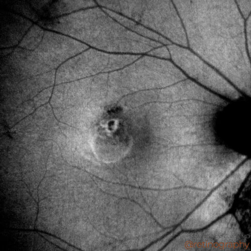 FAF-Green
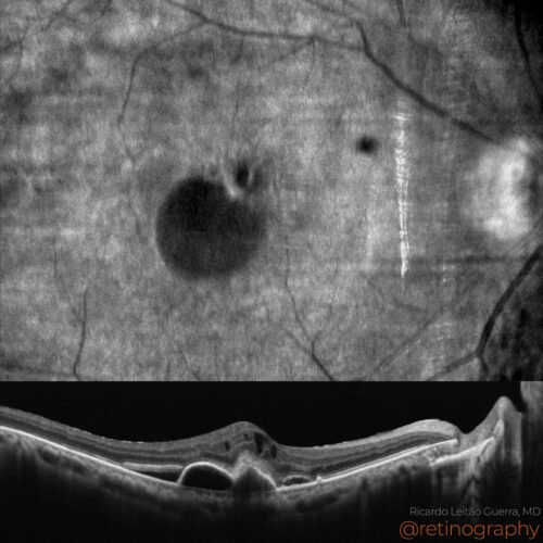 NIR & SD-OCT
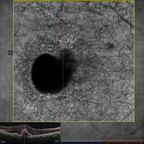 En-face OCT: Structure
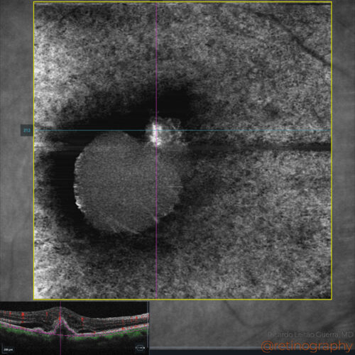 En-face OCT:Angiography
Aneurysmal type 1 neovascularization refers to a subtype of choroidal neovascularization characterized by polypoidal lesions. It is typically identified using OCT and ICGA, showing branching...
Aneurysmal type 1 neovascularization refers to a subtype of choroidal neovascularization characterized by polypoidal lesions. It is typically identified using OCT and ICGA, showing branching...
Aneurysmal type 1 neovascularization refers to a subtype of choroidal neovascularization characterized by polypoidal lesions. It is typically identified using OCT and ICGA, showing branching vascular networks with aneurysmal dilations. This condition may require anti-VEGF therapy and sometimes photodynamic therapy, particularly in treatment-resistant cases.
Disclosure: All images featured in this post were acquired and analyzed using devices integrated within the Zeiss Retina Workflow. This ensures high-quality, detailed visual data for comprehensive assessment.
BackRead More