


Tag:
Type 3 Macular neovascularization
-
Not just drusen – Type 3 MNV
Ricardo Leitão Guerra, MD 74yo
74yo  FAF-Green
 NIR & SD-OCT
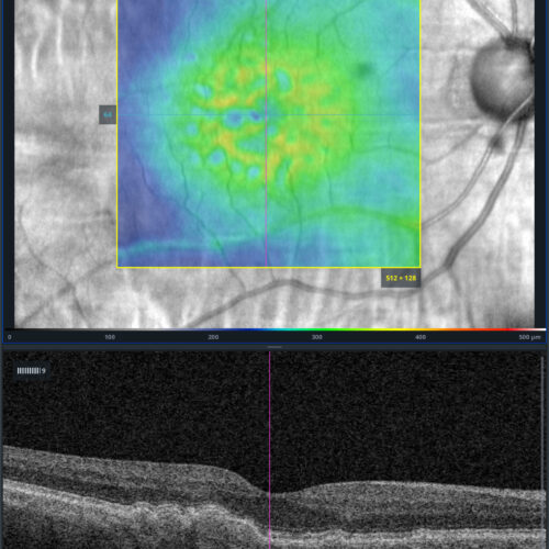 Screenshot
 OCT-Angiography
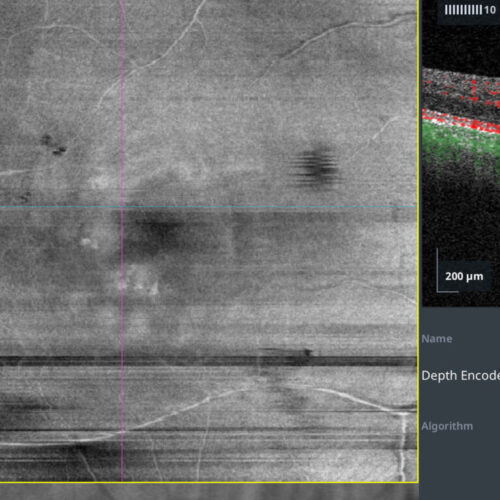 OCT-Angiography
Check out this case: A 73-year-old male presented with apparent non-neovascular age-related macular degeneration (AMD) in the right eye. True color imaging revealed multiple confluent soft d...
Check out this case: A 73-year-old male presented with apparent non-neovascular age-related macular degeneration (AMD) in the right eye. True color imaging revealed multiple confluent soft d...
Check out this case: A 73-year-old male presented with apparent non-neovascular age-related macular degeneration (AMD) in the right eye. True color imaging revealed multiple confluent soft drusen across the macular area. FAF-green imaging showed hypoautofluorescent spots over some of these drusen. SD-OCT B-scan images clearly depicted the drusen as homogeneous, medium-to-high reflectivity elevations of the retinal pigment epithelium (RPE). Advanced RPE analysis enabled precise evaluation of the area and volume of these drusen, facilitating intuitive comparisons over time. However, a more detailed analysis using OCTA revealed type 3 macular neovascularization with adjacent subtle intraretinal fluid. This case underscores the importance of thorough evaluation in AMD patients, utilizing all tools in multimodal retinal imaging for the early detection of more severe forms.
#retina #oftalmo #ophthalmology #oftalmologia #oftalmología #ophtalmologie #офтальмологія #офтальмология #οφθαλμολογία #retinography2024 #CIRRUS6000 #CLARUS700 #ZEISSRETINAWORKFLOW
BackRead More -
Type 3 Macular Neovascularization
Ricardo Leitão Guerra, MD 74yo 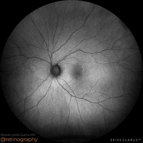 FAF-Green
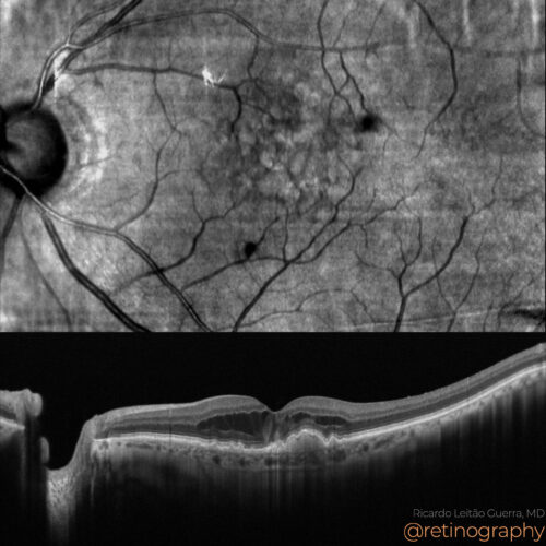 NIR & SD-OCT
 True color: Post Anti-VEGF
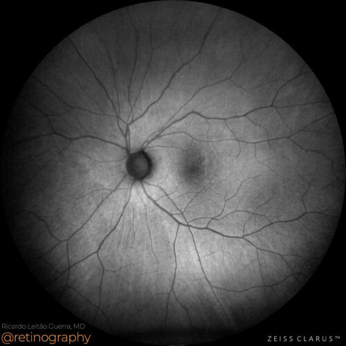 FAF-Green: Post Anti-VEGF
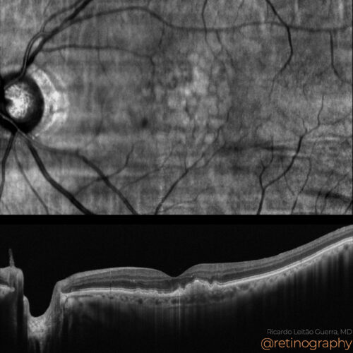 NIR & SD-OCT
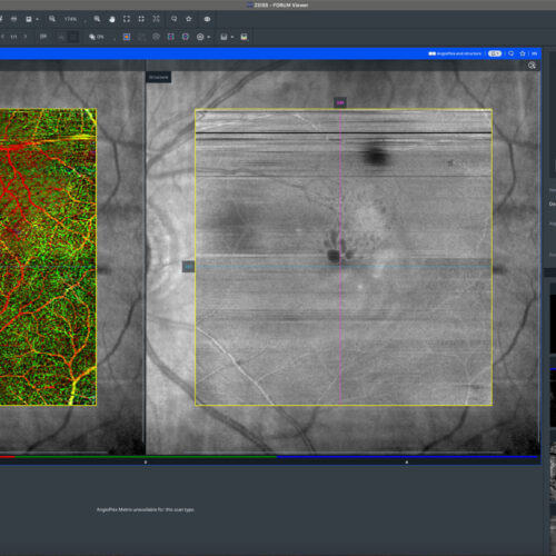 OCT-Angiography
 OCT-Angiography
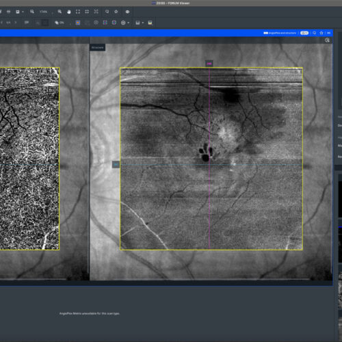 OCT-Angiography
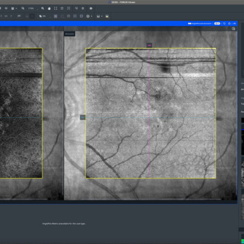 OCT-Angiography
Type 3 macular neovascularization (MNV) in age-related macular degeneration (AMD) is characterized by neovascular growth within the retinal layers. Optical Coherence Tomography Angiography (...
Type 3 macular neovascularization (MNV) in age-related macular degeneration (AMD) is characterized by neovascular growth within the retinal layers. Optical Coherence Tomography Angiography (...
Type 3 macular neovascularization (MNV) in age-related macular degeneration (AMD) is characterized by neovascular growth within the retinal layers. Optical Coherence Tomography Angiography (OCTA) effectively visualizes these intraretinal neovascular networks. Anti-VEGF therapy is the primary treatment, reducing neovascular activity and associated fluid to preserve vision.
#Type3MNV #AMD #OCTA #AntiVEGF #retina #oftalmo #ophthalmology #oftalmologia #oftalmología #ophtalmologie #офтальмологія #офтальмология #οφθαλμολογία #retinography2024 #CIRRUS6000 #CLARUS700 #ZEISSRETINAWORKFLOW
BackRead More -
AMD: Type 3 MNV
Ricardo Leitão Guerra, MD 73yo 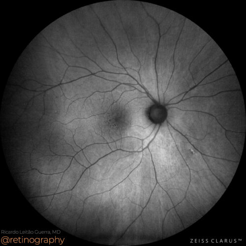 FAF-Green
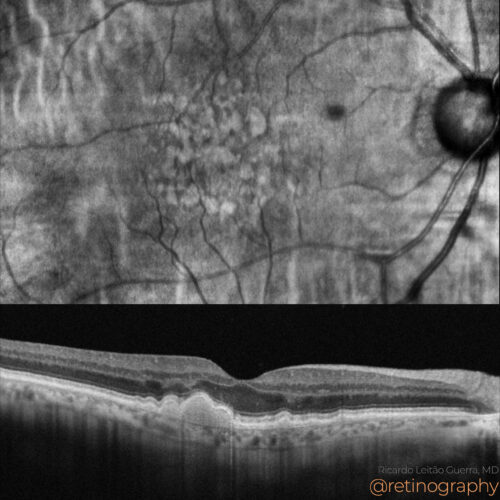 NIR & SD-OCT
 OCT-Angiography
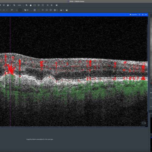 OCT-Angiography
Type 3 macular neovascularization (MNV) is characterized by neovascular growth originating from the deep capillary plexus, extending into the retinal layers. This subtype, also known as reti...
Type 3 macular neovascularization (MNV) is characterized by neovascular growth originating from the deep capillary plexus, extending into the retinal layers. This subtype, also known as reti...
Type 3 macular neovascularization (MNV) is characterized by neovascular growth originating from the deep capillary plexus, extending into the retinal layers. This subtype, also known as retinal angiomatous proliferation (RAP), can be effectively visualized using Optical Coherence Tomography Angiography (OCTA). OCTA provides detailed imaging of the retinal and choroidal microvasculature, facilitating the detection of abnormal blood flow. Intraretinal fluid is commonly associated with Type 3 MNV and can indicate disease progression or response to treatment.
#retina #oftalmo #ophthalmology #oftalmologia #oftalmología #ophtalmologie #офтальмологія #офтальмология #οφθαλμολογία #retinography2024 #CIRRUS6000 #CLARUS700 #ZEISSRETINAWORKFLOW
BackRead More