


Tag:
Vitrectomy
-
Persistent diabetic macular edema
Ricardo Leitão Guerra, MD 75yo
75yo 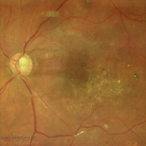 True color: Follow up
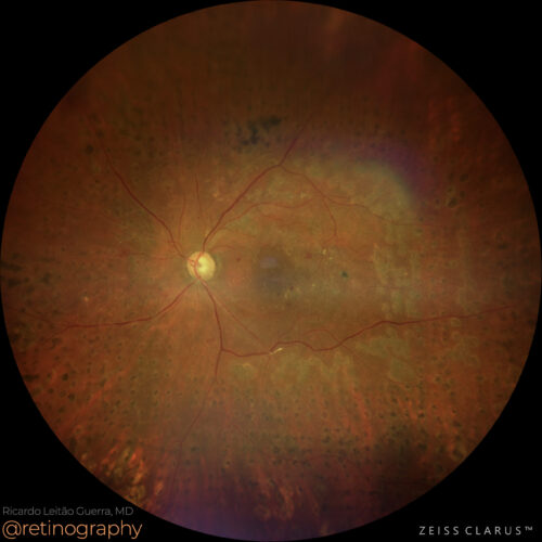 True color - Zoom
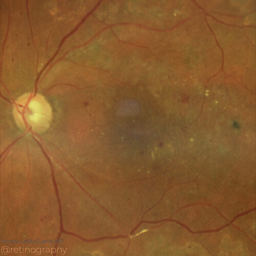 True color - Zoom
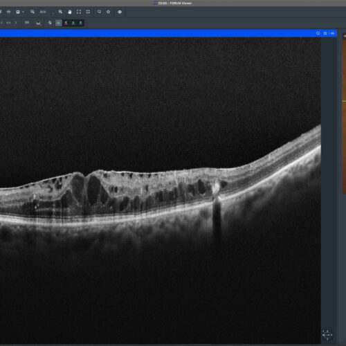 SD-OCT
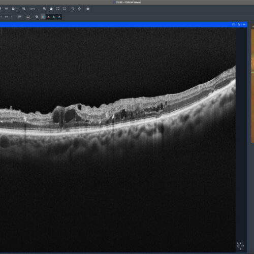 SD-OCT follow up
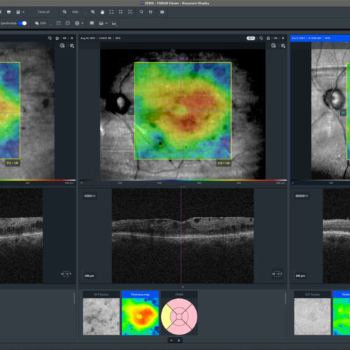 Evolution: Thickness map
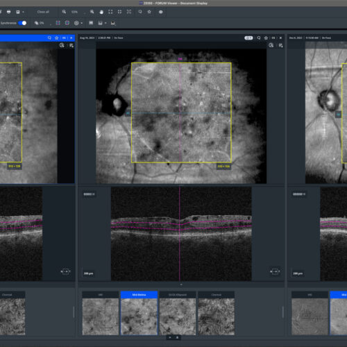 Evolution: En-face
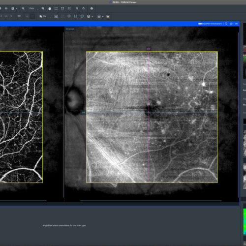 OCT-Angiography
Persistent diabetic macular edema (DME) despite intravitreal anti-VEGF and corticosteroids can be due to traction caused by an epiretinal membrane (ERM). Surgical intervention, such as vitre...
Persistent diabetic macular edema (DME) despite intravitreal anti-VEGF and corticosteroids can be due to traction caused by an epiretinal membrane (ERM). Surgical intervention, such as vitre...
BackRead More -
Epiretinal membrane
Ricardo Leitão Guerra, MD 69yo 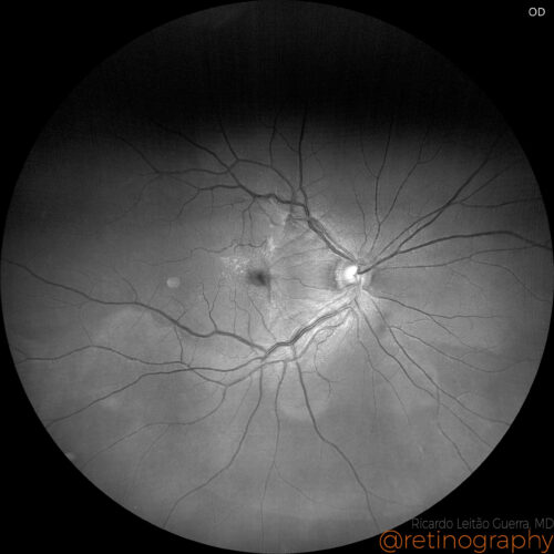 Blue channel - Baseline
 True Color - Postoperative
 Blue channel - Postoperative
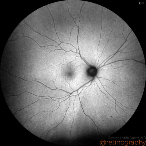 FAF-Green - Postoperative
A beautiful case of an epiretinal membrane peeled from the retina surface. Blue channel allows an enhanced visualization of the inner retinal layers allowing a better surgery planning....
A beautiful case of an epiretinal membrane peeled from the retina surface. Blue channel allows an enhanced visualization of the inner retinal layers allowing a better surgery planning....
A beautiful case of an epiretinal membrane peeled from the retina surface. Blue channel allows an enhanced visualization of the inner retinal layers allowing a better surgery planning.
BackRead More -
Rhegmatogenous Retinal Detachment
Ricardo Leitão Guerra, MD 41yo
41yo  Detached retina - Superior tear
 Retina reattached after PPV + C3F8
A 41yo male presented with rhegmatogenous retinal detachment (macula on) and multiple retinal tears, the larger one was superior. He was treated by primary pars plana vitrectomy. Forty days ...
A 41yo male presented with rhegmatogenous retinal detachment (macula on) and multiple retinal tears, the larger one was superior. He was treated by primary pars plana vitrectomy. Forty days ...
A 41yo male presented with rhegmatogenous retinal detachment (macula on) and multiple retinal tears, the larger one was superior. He was treated by primary pars plana vitrectomy. Forty days after surgery BCVA was 20/20 and C3F8 was filling 1/3 of the vitreous cavity. Vitrectomy with gas injection is a common treatment for retinal detachment, involving the removal of the vitreous gel and injecting a gas bubble to press the retina back into place. The gas bubble helps flatten the retina against the wall of the eye, facilitating reattachment and healing.
Disclosure: All images featured in this post were acquired and analyzed using devices integrated within the Zeiss Retina Workflow. This ensures high-quality, detailed visual data for comprehensive assessment.
BackRead More -
Diabetic tractional retinal detachment
Ricardo Leitão Guerra, MD 46yo  45 days after surgery
True color fundus images in a case os tractional retinal detachment due to proliferative diabetic retinopathy treated with pars plana vitrectomy. Disclosure: All images featured in this post...
True color fundus images in a case os tractional retinal detachment due to proliferative diabetic retinopathy treated with pars plana vitrectomy. Disclosure: All images featured in this post...
True color fundus images in a case os tractional retinal detachment due to proliferative diabetic retinopathy treated with pars plana vitrectomy.
Disclosure: All images featured in this post were acquired and analyzed using devices integrated within the Zeiss Retina Workflow. This ensures high-quality, detailed visual data for comprehensive assessment.
BackRead More -
Retained lens fragments
Ricardo Leitão Guerra, MD 76 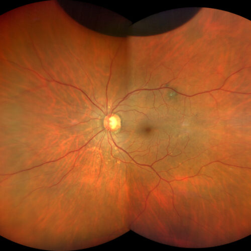 Appearance after vitrectomy - Gas bubble superior
A 76 yo female presented posterior capsule tear and retained lens fragments during phacoemulsification. Vitrectomy was performed 3 days later and final BCVA was 20/20....
A 76 yo female presented posterior capsule tear and retained lens fragments during phacoemulsification. Vitrectomy was performed 3 days later and final BCVA was 20/20....
A 76 yo female presented posterior capsule tear and retained lens fragments during phacoemulsification. Vitrectomy was performed 3 days later and final BCVA was 20/20.
BackRead More